Skip to main content Skip to navigation
Recombinant Human p53 Protein (Wildtype)

BD Pharmingen™ Recombinant Human p53 Protein (Wildtype)

(RUO)
Recombinant Human p53 Protein (Wildtype)
Analysis of purified recombinant p53. Lane 1, Coomassie Blue stained 4-20% mini SDS/PAGE. Lanes 2 and 3, Western blot analysis of p53 using clone PAb 240 (Cat. No. 554166) or a mouse IgG1 isotype (negative) control. A major p53 band is identified at 53 kDa. A lower molecular weight doublet, reactive with anti-p53 antibody, is uncharacterized. 1 µg of purified p53 was loaded into each lane of the gel. MW, Coomassie Blue stained molecular weight standards.
Recombinant Human p53 Protein (Wildtype)
Western blot analysis of recombinant p53 using a panel of antibodies. 150 ng of recombinant protein per lane of a mini gel was separated by SDS/PAGE and detected by Western blotting using p53 monoclonal antibodies that recognize different epitopes. A mouse IgG1 isotype was used as a negative control (Clone A85-1, Cat. No. 553440). The DO-1 and DO-7 antibodies identified a single band at 53 kDa. The other p53 antibodies identified a band at 53 kDa, as well as a lower uncharacterized band which may be a p53 breakdown product. Lane 1: G59-12, lane 2:  PAb 122, lane 3: PAb240, lane 4: DO-1, lane 5: DO-7, lane 6: PAb 1801.
Recombinant Human p53 Protein (Wildtype)
Western blot analysis of a p53 titration. Decreasing amounts of recombinant human p53 were loaded onto lanes of a mini gel separated by SDS/PAGE and detected by Western blotting using a monoclonal antibody to human p53 (clone DO-7, Cat. No. 554294). Protein loaded per lane ranged from 600-19 ng [1, 600 ng), 2 (300 ng), 3 (150 ng), 4 (75 ng), 5 (38 ng), and 6 (19 ng)].
Analysis of purified recombinant p53. Lane 1, Coomassie Blue stained 4-20% mini SDS/PAGE. Lanes 2 and 3, Western blot analysis of p53 using clone PAb 240 (Cat. No. 554166) or a mouse IgG1 isotype (negative) control. A major p53 band is identified at 53 kDa. A lower molecular weight doublet, reactive with anti-p53 antibody, is uncharacterized. 1 µg of purified p53 was loaded into each lane of the gel. MW, Coomassie Blue stained molecular weight standards.
Western blot analysis of recombinant p53 using a panel of antibodies. 150 ng of recombinant protein per lane of a mini gel was separated by SDS/PAGE and detected by Western blotting using p53 monoclonal antibodies that recognize different epitopes. A mouse IgG1 isotype was used as a negative control (Clone A85-1, Cat. No. 553440). The DO-1 and DO-7 antibodies identified a single band at 53 kDa. The other p53 antibodies identified a band at 53 kDa, as well as a lower uncharacterized band which may be a p53 breakdown product. Lane 1: G59-12, lane 2:  PAb 122, lane 3: PAb240, lane 4: DO-1, lane 5: DO-7, lane 6: PAb 1801.
Western blot analysis of a p53 titration. Decreasing amounts of recombinant human p53 were loaded onto lanes of a mini gel separated by SDS/PAGE and detected by Western blotting using a monoclonal antibody to human p53 (clone DO-7, Cat. No. 554294). Protein loaded per lane ranged from 600-19 ng [1, 600 ng), 2 (300 ng), 3 (150 ng), 4 (75 ng), 5 (38 ng), and 6 (19 ng)].
Product Details
Down Arrow Up Arrow


Description

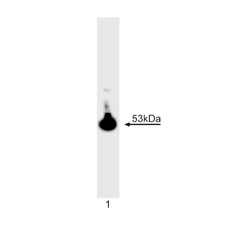
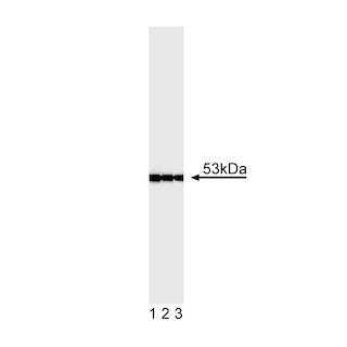
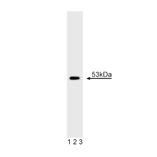
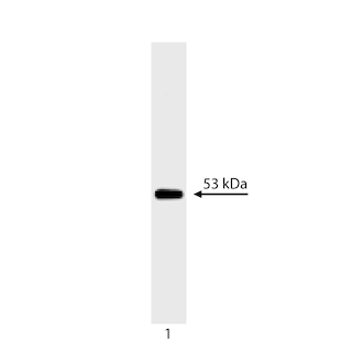
p53 is a nuclear phosphoprotein that plays a key role in cell growth regulation, particularly inhibition of cell proliferation. BD Biosciences Pharmingen offers a panel of p53 specific antibodies. The recombinant p53 protein is useful as a western blot control for antibodies that recognize human p53 and work in western blot. These antibodies include the following p53 clones: G59-12, PAb 122, PAb 240, PAb 1801, DO-1, DO-7. Recombinant human p53 was expressed in the Baculovirus Expression Vector System (BEVS) and purified from Sf9 insect cell lysate using an anti-p53 antibody, clone DO-7 (Cat. No. 554294).

The purity and the integrity of the protein was verified by SDS-PAGE and Western blot analysis.  


Preparation And Storage

This preparation contains no preservatives, thus it should be handled under aseptic conditions. Avoid multiple freeze-thaws of product.

The protein solution was sterile filtered and frozen at -20°C. Store the protein at -20°C; it may also be stored at -80°C.

Recommended Assay Procedures

1 µg of protein can easily be visualized on a Coomassie Blue stained SDS/PAGE as a band of 53 kDa. For Western blot analysis, 50 ng or less is sufficient for the chemiluminescence detection method. The p53 recombinant protein has not been evaluated for suitability in other assays outside of Western blot analysis.

Product Notices

  1. Since applications vary, each investigator should titrate the reagent to obtain optimal results.
  2. Please refer to www.bdbiosciences.com/us/s/resources for technical protocols.
  3. Source of all serum proteins is from USDA inspected abattoirs located in the United States.
556439 Rev. 7

Please refer to Support Documents for Quality Certificates

Global - Refer to manufacturer's instructions for use and related User Manuals and Technical data sheets before using this products as described

Comparisons, where applicable, are made against older BD Technology, manual methods or are general performance claims.  Comparisons are not made against non-BD technologies, unless otherwise noted.

For Research Use Only. Not for use in diagnostic or therapeutic procedures.

Refer to manufacturer's instructions for use and related User Manuals and Technical Data Sheets before using this product as described.

Comparisons, where applicable, are made against older BD technology, manual methods or are general performance claims. Comparisons are not made against non-BD technologies, unless otherwise noted.