Preparation And Storage
1. For in vitro diagnostic use.
2. When stored at 2° to 8°C, the antibody reagent is stable until the expiration date shown on the label. Do not use after the expiration date.
3. The antibody reagent should not be frozen or exposed to direct light during storage or during incubation with cells. Keep the reagent vial dry.
4. Alteration in the appearance of the reagent, such as precipitation or discoloration, indicates instability or deterioration. In such cases, the reagent should not be used.
5. The antibody reagent contains sodium azide as a preservative; however, care should be taken to avoid microbial contamination, which may cause erroneous results.
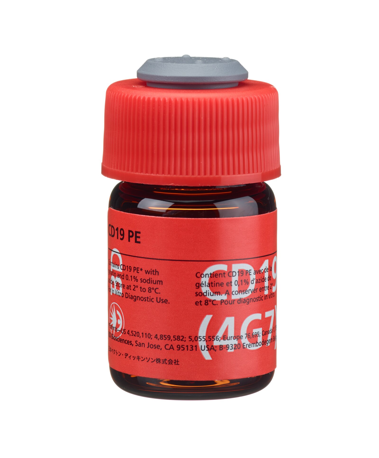
Becton Dickinson Immunocytometry Systems (BDIS) CD19 (Leu™-12) phycoerythrin (PE) is a single-color direct immunofluorescence reagent for the enumeration of B lymphocytes (CD19+) in peripheral blood using fluorescence microscopy or flow cytometers such as the FACSCalibur™, FACSort™, FACScan™, or FACS Analyzer™.

Development References (20)
-
Brown G, Greaves M. Enumeration of absolute numbers of T and B cells in human blood. Scand J Immunol. 1974; 3:161. (Biology).
-
Centers for Disease Control. Guidelines for the performance of CD4+ T-cell determination in persons with human immunodeficiency virus infection. MMWR. 1992; 41:1-17. (Biology).
-
Centers for Disease Control. Update: universal precautions for prevention of transmission of humanimmunodeficiency virus, hepatitis B virus, and other bloodborne pathogens in health-care settings. MMWR. 1988; 37:377-388. (Biology).
-
Clark P, Normansell D, Innes D, Hess C. Lymphocyte subsets in normal bone marrow. Blood. 1986; 67:1600. (Biology).
-
Dwyer J, Finklestein F, Mangi R, Fisher K, Hendler E. Assessment of the adequacy of immunosuppressive therapy using microscopy techniques to study immunologic competence. Transplant Proc. 1975; 7:785. (Biology).
-
Dörken B, Möller P, Pezzutto A, Schwartz-Albiez R, Moldenhauer G. Knapp W, Dörken B, Gilks WR, et al, ed. Leucocyte Typing IV: White Cell Differentiation Antigens. New York, NY: Oxford University Press; 1989:34-36.
-
Foucar K, Goeken JA. Clinical application of immunologic techniques to the diagnosis of lymphoproliferative and immunodeficiency disorders. Lab Med. 1982; 13:403-413. (Biology).
-
Fröland S, Natvig J, Berdal P Surface-bound immunoglobulin as a marker of B lymphocytes in man. Nature. 1971; 251-252. (Biology).
-
Giorgi JV. Lymphocyte subset measurements: significance in clinical medicine. In: Rose NR, Friedman H, Fahey JL, ed. Manual of Clinical Laboratory Immunology. 3rd ed.. Washington, DC: American Society for Microbiology; 1986:236-246.
-
Jackson AL, Warner NL. Rose NR, Friedman H, Fahey JL, ed. Manual of Clincial Laboratory Immunology, Third Edition. Washington DC: American Society for Microbiology; 1986:226-235.
-
Kohler G, Milstein C. Continuous cultures of fused cells secreting antibody of predefined specificity. Nature. 1975; 256:495. (Biology).
-
Loken MR, Shah VO, Dattilio KL, Civin CI. Flow cytometric analysis of human bone marrow. II. Normal B lymphocyte development. Blood. 1987; 70(5):1316-1324. (Biology). View Reference
-
Meeker TC, Miller RA, Link MP, Bindl J, Warnke R, Levy R. A unique human B lymphocyte antigen defined by a monoclonal antibody.. Hybridoma. 1984; 3(4):305-20. (Biology). View Reference
-
Mishell B, Shiigi S, Henry C, et al. Mishell B, Shiigi S, ed. Selected Methods in Cellular Immunology. New York: WH Freeman and Co; 1980:16-17.
-
Nadler LM. B Cell/Leukemia Panel Workshop: summary and comments. In: Reinherz EL, Haynes BF, Nadler LM, Bernstein ID, ed. Leukocyte Typing II: Human B Lymphocytes. New York: Springer-Verlag; 1986:3-43.
-
National Committee for Clinical Laboratory Standards. Clinical Applications of Flow Cytometry: Quality Assurance and Immunophenotyping of Peripheral Blood Lymphocytes; Tentative Guideline. 1992; 1992. (Biology).
-
National Committee for Clinical Laboratory Standards. Protection of Laboratory Workers from Infectious Disease Transmitted by Blood, Body Fluids, and Tissue: Tentative Guideline (M29-T2). 1991. (Biology).
-
Ryan D, Kossover S, Mitchell S, Frantz C, Hennessy L, Cohen H. Subpopulations of common acute lymphoblastic leukemia antigen-positive lymphoid cells in normal bone marrow identified by hematopoietic differentiation antigens.. Blood. 1986; 68(2):417-25. (Biology). View Reference
-
Tursz T, Brouet J-C, Flandrin G, Danon F, Clauvel J-P, Seligmann M. Clinical and pathologic features of Waldenström's macroglobulinemia in seven patients with serum monoclonal IgG or IgA. Am J Med. 1977; 63:499-502. (Biology).
-
Warner N. Membrane immunoglobulins and antigen receptors on B and T lymphocytes. Advances Immunol. 1974; 19:67. (Biology).
Please refer to Support Documents for Quality Certificates
Global - Refer to manufacturer's instructions for use and related User Manuals and Technical data sheets before using this products as described
Comparisons, where applicable, are made against older BD Technology, manual methods or are general performance claims. Comparisons are not made against non-BD technologies, unless otherwise noted.
For In Vitro Diagnostics Use.
Refer to manufacturer's instructions for use and related User Manuals and Technical Data Sheets before using this product as described.
Comparisons, where applicable, are made against older BD technology, manual methods or are general performance claims. Comparisons are not made against non-BD technologies, unless otherwise noted.