-
Training
- Flow Cytometry Basic Training
-
Product-Based Training
- BD FACSDiscover™ S8 Cell Sorter Product Training
- Accuri C6 Plus Product-Based Training
- FACSAria Product Based Training
- FACSCanto Product-Based Training
- FACSLyric Product-Based Training
- FACSMelody Product-Based Training
- FACSymphony Product-Based Training
- HTS Product-Based Training
- LSRFortessa Product-Based Training
- Advanced Training
-
- BD FACSDiscover™ S8 Cell Sorter Product Training
- Accuri C6 Plus Product-Based Training
- FACSAria Product Based Training
- FACSCanto Product-Based Training
- FACSLyric Product-Based Training
- FACSMelody Product-Based Training
- FACSymphony Product-Based Training
- HTS Product-Based Training
- LSRFortessa Product-Based Training
- United States (English)
-
Change country/language
Old Browser
This page has been recently translated and is available in French now.
Looks like you're visiting us from {countryName}.
Would you like to stay on the current country site or be switched to your country?


Western blot analysis of ERK2 on rat pituitary lysate. Lane 1: 1:5000, lane 2: 1:10000, lane 3: 1:20000 dilution of ERK2.

ERK 2 (clone 33) staining on rat brain. Formalin fixed paraffin section with citrate buffer pretreatment. 20X

Immunofluorescence staining of MCF7 cells.


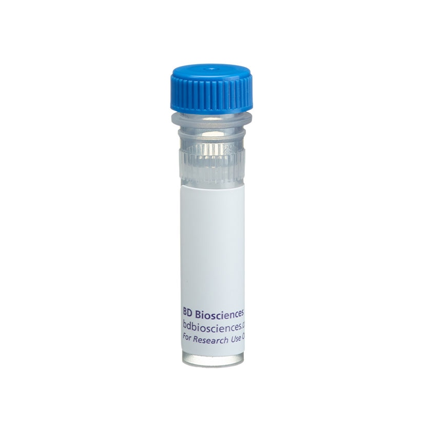
BD Transduction Laboratories™ Purified Mouse Anti-ERK2

BD Transduction Laboratories™ Purified Mouse Anti-ERK2

BD Transduction Laboratories™ Purified Mouse Anti-ERK2

Regulatory Status Legend
Any use of products other than the permitted use without the express written authorization of Becton, Dickinson and Company is strictly prohibited.
Preparation And Storage
Product Notices
- Since applications vary, each investigator should titrate the reagent to obtain optimal results.
- Please refer to www.bdbiosciences.com/us/s/resources for technical protocols.
- Caution: Sodium azide yields highly toxic hydrazoic acid under acidic conditions. Dilute azide compounds in running water before discarding to avoid accumulation of potentially explosive deposits in plumbing.
- Source of all serum proteins is from USDA inspected abattoirs located in the United States.
Companion Products

.png?imwidth=320)
The family of serine/threonine kinases known as ERKs (extracellular signal regulated kinases) or MAPKs (mitogen-activated protein kinases) are activated after cell stimulation by a wide variety of hormones and growth factors. Cell stimulation induces a signaling cascade that leads to phosphorylation of MEK (MAPK/ERK kinase) which, in turn, activates ERK via tyrosine and threonine phosphorylation. Structural analysis of ERK2 indicates that phosphorylation induces a conformational change that exposes the active site for substrate binding. Myriad proteins represent the downstream effectors for the active ERK and implicate it in the control of cell proliferation and differentiation, as well as regulation of the cytoskeleton. Activation of ERK is normally transient and cells possess dual specificity phosphatases that are responsible for its down-regulation. Furthermore, multiple studies have shown that elevated ERK activity is associated with some cancers. ERK2 is the 42kDa member of the ERK family and is highly homologous to ERK1.
Development References (5)
-
Kim SJ, Ju JW, Oh CD. ERK-1/2 and p38 kinase oppositely regulate nitric oxide-induced apoptosis of chondrocytes in association with p53, caspase-3, and differentiation status. J Biol Chem. 2002; 277(2):1332-1339. (Clone-specific: Western blot). View Reference
-
Lehmann K, Janda E, Pierreux CE, et al. Raf induces TGFbeta production while blocking its apoptotic but not invasive responses: a mechanism leading to increased malignancy in epithelial cells. Genes Dev. 2000; 14(20):2610-2622. (Clone-specific: Western blot). View Reference
-
Liu L, Tsai JC, Aird WC. Egr-1 gene is induced by the systemic administration of the vascular endothelial growth factor and the epidermal growth factor. Blood. 1772; 96(5):1772-1781. (Clone-specific: Western blot). View Reference
-
Lund-Johansen F, Davis K, Bishop J, de Waal Malefyt R. Flow cytometric analysis of immunoprecipitates: high-throughput analysis of protein phosphorylation and protein-protein interactions. Cytometry. 2000; 39(4):250-259. (Clone-specific: Flow cytometry, Immunoprecipitation, Western blot). View Reference
-
Visconti R, Gadina M, Chiariello M. Importance of the MKK6/p38 pathway for interleukin-12-induced STAT4 serine phosphorylation and transcriptional activity. Blood. 2000; 96(5):1844-1852. (Clone-specific: Immunoprecipitation, In vitro kinase assay, Western blot). View Reference
Please refer to Support Documents for Quality Certificates
Global - Refer to manufacturer's instructions for use and related User Manuals and Technical data sheets before using this products as described
Comparisons, where applicable, are made against older BD Technology, manual methods or are general performance claims. Comparisons are not made against non-BD technologies, unless otherwise noted.
For Research Use Only. Not for use in diagnostic or therapeutic procedures.
Report a Site Issue
This form is intended to help us improve our website experience. For other support, please visit our Contact Us page.