Skip to main content Skip to navigation
Purified Rabbit Anti- Active Caspase-3
Alert icon
This product will be discontinued on June 2024. Consider alternate product [570524], clone C92-605.rMAb, which is manufactured with recombinant technology
Purified Rabbit Anti- Active Caspase-3
Immunoprecipitation/Western blot analysis of caspase-3 from apoptotic and non-apoptotic cell lysates. Jurkat cells (Human T-cell leukemia; ATCC TIB-152) were left untreated (left panel) or were treated with 6 µM camptothecin for 5 hr to induce apoptosis (right panel). Immunoprecipitation: Cell lysates were immunoprecipitated with 0.25 - 2 µg/ml of the rabbit anti- active caspase-3 antibody [clone C92-605, lanes 1 and 8 (2 µg), lanes 2 and 9, (1 µg), lanes 3 and 10 (0.5 µg), and lanes 4 and 11 (0.25 µg)], 1 - 2 µg/ml of a mAb recognizing both pro and active caspase-3 (Cat. No. 610322), lanes 5 and 12 (2 µg), lanes 6 and 13 (1 µg)] or 1 µg/ml of a rabbit IgG isotype control [Jackson Immunoresearch (Cat. No.  011-00000-3), lanes 7 and 14]. Western blot: Caspase-3 was detected by western blot analysis with an antibody that recognizes both pro- (32 kD) and active (20 and 17 kD, reflecting the presence or absence of the caspase-3 pro-domain) caspase-3 [lanes 1-14]. The results also show that the rabbit anti- active caspase-3 antibody (clone C92-605) immunoprecipitated only the active form of caspase-3 (lanes 8-11) as compared to the mouse anti-human caspase-3 antibody (Cat. No. 610322) which immunoprecipitated both the pro and active forms of caspase-3 (lanes 12 and 13). Bands may be observable at ~25 kD and ~55-60 kD which represent the light and heavy chains, respectively, of IgG used for the immunoprecipitation.
Product Details
Down Arrow Up Arrow


BD Pharmingen™
CPP32; Yama; Apopain
Human (QC Testing), Mouse (Tested in Development)
Rabbit IgG
Human Active Caspase-3 Fragment
Immunoprecipitation/Western blot, Intracellular staining (flow cytometry) (Routinely Tested), Bioimaging, ELISA, Immunohistochemistry-frozen (Tested During Development)
20 kD & 17 kD
0.5 mg/ml
AB_397274
Aqueous buffered solution containing ≤0.09% sodium azide.
RUO


Preparation And Storage

Store undiluted at 4°C. The monoclonal antibody was purified from tissue culture supernatant or ascites by affinity chromatography.

Recommended Assay Procedures

Bioimaging:  Please refer to http://www.bdbiosciences.com/support/resources/bioimaging/index.jsp

Product Notices

  1. Since applications vary, each investigator should titrate the reagent to obtain optimal results.
  2. Please refer to www.bdbiosciences.com/us/s/resources for technical protocols.
  3. Caution: Sodium azide yields highly toxic hydrazoic acid under acidic conditions. Dilute azide compounds in running water before discarding to avoid accumulation of potentially explosive deposits in plumbing.
559565 Rev. 11
Antibody Details
Down Arrow Up Arrow
C92-605

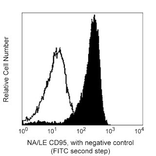
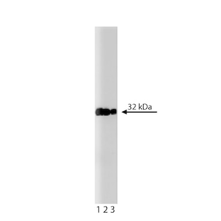
The caspase family of cysteine proteases plays a key role in apoptosis and inflammation. Caspase-3 is a key protease that is activated during the early stages of apoptosis and, like other members of the caspase family, is synthesized as an inactive pro-enzyme that is processed in cells undergoing apoptosis by self-proteolysis and/or cleavage by another protease. The processed forms of caspases consist of large (17-22 kDa) and small (10-12 kDa) subunits which associate to form an active enzyme. Active caspase-3, a marker for cells undergoing apoptosis, consists of a heterodimer of 17 and 12 kDa subunits which is derived from the 32 kDa pro-enzyme. Active caspase-3 proteolytically cleaves and activates other caspases, as well as relevant targets in the cytoplasm, e.g., D4-GDI and Bcl-2, and in the nucleus (e.g. PARP).  This antibody has been reported to specifically recognize the active form of caspase-3 in human and mouse cells.  It has not been reported to recognize the pro-enzyme form of caspase-3.

559565 Rev. 11
Format Details
Down Arrow Up Arrow
Purified
Tissue culture supernatant is purified by either protein A/G or affinity purification methods. Both methods yield antibody in solution that is free of most other soluble proteins, lipids, etc. This format provides pure antibody that is suitable for a number of downstream applications including: secondary labeling for flow cytometry or microscopy, ELISA, Western blot, etc.
Purified
559565 Rev.11
Citations & References
Down Arrow Up Arrow

Development References (3)

  1. Dai C, Krantz SB. Interferon gamma induces upregulation and activation of caspases 1, 3, and 8 to produce apoptosis in human erythroid progenitor cells. Blood. 1999; 93(10):3309-3316. (Biology). View Reference
  2. Fujita N, Tsuruo T. Involvement of Bcl-2 cleavage in the acceleration of VP-16-induced U937 cell apoptosis. Biochem Biophys Res Commun. 1998; 246(2):484-488. (Biology). View Reference
  3. Thornberry NA, Lazebnik Y. Caspases: enemies within. Science. 1998; 281(5381):1312-1316. (Biology). View Reference
559565 Rev. 11

Please refer to Support Documents for Quality Certificates


Global - Refer to manufacturer's instructions for use and related User Manuals and Technical data sheets before using this products as described


Comparisons, where applicable, are made against older BD Technology, manual methods or are general performance claims.  Comparisons are not made against non-BD technologies, unless otherwise noted.

For Research Use Only. Not for use in diagnostic or therapeutic procedures.