
Preparation And Storage
Recommended Assay Procedures
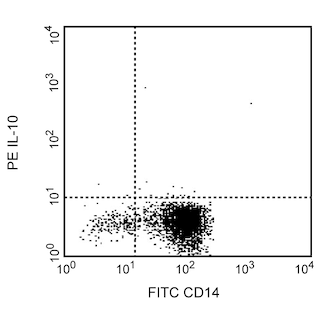
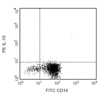
1. Immunofluorescent Staining and Flow Cytometric Analysis: The PE-conjugated JES3-19F1 antibody (Cat. No. 554706) can be used for multicolor immunofluorescent staining and flow cytometric analyses to identify and enumerate IL-10-producing cells within mixed cell populations (see image). For optimal immunofluorescent staining with flow cytometric analysis, this anti-cytokine antibody should be titrated (≤ 0.5 µg mAb/million cells).
A useful control for demonstrating specificity of staining is either of the following: 1) pre-block the conjugated JES3-19F1 antibody with ligand (e.g., recombinant human IL-10; Cat. No. 554611) prior to staining, or 2) pre-block the fixed/ permeabilized cells with unlabelled JES3-19F1 antibody (Cat. No. 554705) prior to staining. The staining technique and use of blocking controls are described in detail by C. Prussin and D. Metcalfe. A suitable rat IgG2a isotype control for assessing the level of background staining on paraformaldehyde-fixed/saponin-permeabilized human cells is PE-R35-95 (Cat. No. 554689); use at comparable concentrations to the antibody of interest (e.g., ≤ 0.5 µg mAb/1 million cells).
BD™ CompBeads can be used as surrogates to assess fluorescence spillover (Compensation). When fluorochrome conjugated antibodies are bound to CompBeads, they have spectral properties very similar to cells. However, for some fluorochromes there can be small differences in spectral emissions compared to cells, resulting in spillover values that differ when compared to biological controls. It is strongly recommended that when using a reagent for the first time, users compare the spillover on cell and CompBead to ensure that BD Comp beads are appropriate for your specific cellular application.
2. ELISA Capture: The purified JES3-19F1 (Cat. No. 554705) antibody is useful as a capture antibody for a sandwich ELISA for specifically measuring human IL-10 protein levels. Purified JES3-19F1 antibody can be paired with the biotinylated JES3-12G8 antibody (Cat. No. 554499) as the detection antibody and with recombinant human IL-10 protein (Cat. No. 554611) as the standard. For testing IL-10 in serum or plasma, our OptEIA™ set (Cat. No. 555134) is recommended.
3. Neutralization: The NA/LE™ format of clone JES3-19F1 (Cat. No. 554704) is useful for neutralization of human IL-10 bioactivity. A suitable NA/LE rat IgG2a isotype control to match the NA/LE JES3-19F1 antibody is the R35-95 antibody, (Cat. No. 554687).
Product Notices
- Since applications vary, each investigator should titrate the reagent to obtain optimal results.
- An isotype control should be used at the same concentration as the antibody of interest.
- Caution: Sodium azide yields highly toxic hydrazoic acid under acidic conditions. Dilute azide compounds in running water before discarding to avoid accumulation of potentially explosive deposits in plumbing.
- For fluorochrome spectra and suitable instrument settings, please refer to our Multicolor Flow Cytometry web page at www.bdbiosciences.com/colors.
- Species cross-reactivity detected in product development may not have been confirmed on every format and/or application.
- Please refer to http://regdocs.bd.com to access safety data sheets (SDS).
- Please refer to www.bdbiosciences.com/us/s/resources for technical protocols.
Companion Products
.png?imwidth=320)
The JES3-19F1 monoclonal antibody specifically recognizes human Interleukin-10 (IL-10) that is encoded by IL10. IL-10 is also known as Cytokine Synthesis Inhibitory Factor (CSIF), B cell-derived T cell growth factor (B-TCGF), and T-cell growth inhibitory factor (TGIF). The JES3-19F1 antibody crossreacts with ebvIL-10 protein, the Epstein-Barr viral IL-10 homolog (viral IL-10 or vIL-10) encoded by the BCRF1 gene. IL-10 is produced by a variety of cells such as some activated T cells and B cells including regulatory T cells (Treg) and B cells (Breg), monocytes and macrophages, dendritic cells (DC), keratinocytes, and mast cells. IL-10 is a multifunctional cytokine that can downregulate immune and proinflammatory responses. IL-10 can act to reduce expression of major histocompatibility complex class II antigens, costimulatory molecules, or proinflammatory cytokines including IL-1β, IL-2, IL-3, IL-12, IFN-γ, TNF or GM-CSF expressed by activated monocytes, macrophages, dendritic cells (DC), natural killer (NK) cells, or T cells. IL-10 has been shown to play a role in chronic viral infections. IL-10 can also enhance B cell survival, proliferation, and differentiation to become antibody-producing cells. The JES3-19F1 antibody reportedly neutralizes the biological activity of human IL-10 and ebvIL-10. IL-10 mediates its biological activities by signaling through a heterotetrameric receptor complex composed of the type II cytokine receptor subunits CD210a (IL-10 Rα) and CD210b (IL-10 Rβ).

Development References (3)
-
Andersson EC, Christensen JP, Marker O, Thomsen AR. Changes in cell adhesion molecule expression on T cells associated with systemic virus infection. Immunology. 1994; 152(3):1237-1245. (Clone-specific: Blocking). View Reference
-
D'Andrea A, Aste-Amezaga M, Valiante NM, Ma X, Kubin M, Trinchieri G. Interleukin 10 (IL-10) inhibits human lymphocyte interferon gamma-production by suppressing natural killer cell stimulatory factor/IL-12 synthesis in accessory cells. J Exp Med. 1993; 178(3):1041-1048. (Clone-specific: Neutralization). View Reference
-
Prussin C, Metcalfe DD. Detection of intracytoplasmic cytokine using flow cytometry and directly conjugated anti-cytokine antibodies. J Immunol Methods. 1995; 188(1):117-128. (Methodology). View Reference
Please refer to Support Documents for Quality Certificates
Global - Refer to manufacturer's instructions for use and related User Manuals and Technical data sheets before using this products as described
Comparisons, where applicable, are made against older BD Technology, manual methods or are general performance claims. Comparisons are not made against non-BD technologies, unless otherwise noted.
For Research Use Only. Not for use in diagnostic or therapeutic procedures.