Skip to main content Skip to navigation
Alexa Fluor® 647 Mouse anti-GFAP
Analysis of GFAP in differentiated human Neural Stem Cells (NSC). NSC derived from H9 cells (WiCell, Madison, WI) were differentiated in NSC differentiation medium [containing N2 and B-27 supplements (Life Technologies), recombinant human BDNF and GDNF (Peprotech), dibutryl cyclic AMP (Sigma)] for 11 days followed by AGM™ Astrocyte Growth Medium (Lonza) for 16 days. The cells were fixed (BD CytofixTM buffer, Cat. No. 554655) for 20 minutes at room temperature, permeabilized with BD™ Phosflow Perm/Wash Buffer I (Cat. No.557885), and then stained with Alexa Fluor® 647 Mouse anti-GFAP (left panel) and co-stained with FITC mouse anti-CD44 (Cat. No. 555478) as shown in the right panel. This antibody conjugate also works with BD ™ Phosflow Perm Buffer III. Flow cytometry was performed on a BD LSR™ II flow cytometer.
Alexa Fluor® 647 Mouse anti-GFAP
Analysis of GFAP in Neural Stem cells (NSC). NSCs were isolated by sorting from Embryoid bodies and were grown for 8 passages post sort, fixed (BD Cytofix™ buffer, Cat. No. 554655) for 20 minutes at room temperature, permeabilized with BD™ Phosflow Perm Buffer I (Cat. No.557885), and then stained with either Alexa Fluor® 647 Mouse anti-GFAP (solid line) or Alexa Fluor® 647 Mouse IgG2b, κ Isotype Control (Cat. No. 558713, dashed line).
Analysis of GFAP in differentiated human Neural Stem Cells (NSC). NSC derived from H9 cells (WiCell, Madison, WI) were differentiated in NSC differentiation medium [containing N2 and B-27 supplements (Life Technologies), recombinant human BDNF and GDNF (Peprotech), dibutryl cyclic AMP (Sigma)] for 11 days followed by AGM™ Astrocyte Growth Medium (Lonza) for 16 days. The cells were fixed (BD CytofixTM buffer, Cat. No. 554655) for 20 minutes at room temperature, permeabilized with BD™ Phosflow Perm/Wash Buffer I (Cat. No.557885), and then stained with Alexa Fluor® 647 Mouse anti-GFAP (left panel) and co-stained with FITC mouse anti-CD44 (Cat. No. 555478) as shown in the right panel. This antibody conjugate also works with BD ™ Phosflow Perm Buffer III. Flow cytometry was performed on a BD LSR™ II flow cytometer.
Analysis of GFAP in Neural Stem cells (NSC). NSCs were isolated by sorting from Embryoid bodies and were grown for 8 passages post sort, fixed (BD Cytofix™ buffer, Cat. No. 554655) for 20 minutes at room temperature, permeabilized with BD™ Phosflow Perm Buffer I (Cat. No.557885), and then stained with either Alexa Fluor® 647 Mouse anti-GFAP (solid line) or Alexa Fluor® 647 Mouse IgG2b, κ Isotype Control (Cat. No. 558713, dashed line).
Product Details
Down Arrow Up Arrow


BD Pharmingen™
Glial Fibrillary Acidic Protein, FLJ45472
Human (QC Testing), Mouse, Rat, Pig, Dog, Chicken, Rabbit, Cow, Guinea Pig, Sheep (Reported)
Mouse IgG2b
Cow spinal cord homogenate
Intracellular staining (flow cytometry) (Routinely Tested)
5 µl
AB_10646037
Aqueous buffered solution containing BSA, protein stabilizer, and ≤0.09% sodium azide.
RUO


Preparation And Storage

The monoclonal antibody was purified from tissue culture supernatant or ascites by affinity chromatography. The antibody was conjugated to Alexa Fluor® 647 under optimum conditions, and unreacted Alexa Fluor® 647 was removed. Store undiluted at 4°C and protected from prolonged exposure to light. Do not freeze.

Recommended Assay Procedures

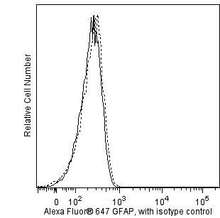
The formulation of this antibody conjugate has been optimized for flow cytometry. We recommend Cat. No. 560298 for Bioimaging.

Product Notices

  1. This reagent has been pre-diluted for use at the recommended Volume per Test. We typically use 1 × 10^6 cells in a 100-µl experimental sample (a test).
  2. Please refer to www.bdbiosciences.com/us/s/resources for technical protocols.
  3. Alexa Fluor® 647 fluorochrome emission is collected at the same instrument settings as for allophycocyanin (APC).
  4. For fluorochrome spectra and suitable instrument settings, please refer to our Multicolor Flow Cytometry web page at www.bdbiosciences.com/colors.
  5. The Alexa Fluor®, Pacific Blue™, and Cascade Blue® dye antibody conjugates in this product are sold under license from Molecular Probes, Inc. for research use only, excluding use in combination with microarrays, or as analyte specific reagents. The Alexa Fluor® dyes (except for Alexa Fluor® 430), Pacific Blue™ dye, and Cascade Blue® dye are covered by pending and issued patents.
  6. Caution: Sodium azide yields highly toxic hydrazoic acid under acidic conditions. Dilute azide compounds in running water before discarding to avoid accumulation of potentially explosive deposits in plumbing.
  7. Source of all serum proteins is from USDA inspected abattoirs located in the United States.
  8. Alexa Fluor® is a registered trademark of Molecular Probes, Inc., Eugene, OR.
561470 Rev. 1
Antibody Details
Down Arrow Up Arrow
1B4

GFAP (Glial Fibrillary Acidic Protein) is the major protein of glial filaments in differentiated astrocytes. BD Biosciences offers a panel of monoclonal antibodies (4A11, 1B4, 2E1) that specifically recognize GFAP. They do not cross-react with other intermediate filaments such as vimentin, neurofilament proteins, desmin, keratin, neurotubules or microfilaments.

561470 Rev. 1
Format Details
Down Arrow Up Arrow
Alexa Fluor™ 647
Alexa Fluor™ 647 Dye is part of the BD red family of dyes. This is a small organic fluorochrome with an excitation maximum (Ex Max) at 653-nm and an emission maximum (Em Max) at 669-nm. Alexa Fluor 647 is designed to be excited by the Red laser (627-640 nm) and detected using an optical filter centered near 520-nm (e.g., a 660/20 nm bandpass filter). Please ensure that your instrument’s configurations (lasers and optical filters) are appropriate for this dye.
Alexa Fluor™ 647
Red 627-640 nm
653 nm
669 nm
561470 Rev.1
Citations & References
Down Arrow Up Arrow

Development References (3)

  1. McLendon RE, Bigner DD. Immunohistochemistry of the glial fibrillary acidic protein: basic and applied considerations. Brain Pathol. 1994; 4(3):221-228. (Biology). View Reference
  2. McLendon RE, Burger PC, Pegram CN, Eng LF, Bigner DD. The immunohistochemical application of three anti-GFAP monoclonal antibodies to formalin-fixed, paraffin-embedded, normal and neoplastic brain tissues. J Neuropathol Exp Neurol. 1986; 45(6):692-703. (Biology). View Reference
  3. Pegram CN, Eng LF, Wikstrand CJ, McComb RD, Lee YL, Bigner DD. Monoclonal antibodies reactive with epitopes restricted to glial fibrillary acidic proteins of several species. Neurochem Pathol. 1985; 3(2):119-138. (Biology). View Reference
561470 Rev. 1

Please refer to Support Documents for Quality Certificates


Global - Refer to manufacturer's instructions for use and related User Manuals and Technical data sheets before using this products as described


Comparisons, where applicable, are made against older BD Technology, manual methods or are general performance claims.  Comparisons are not made against non-BD technologies, unless otherwise noted.

For Research Use Only. Not for use in diagnostic or therapeutic procedures.