-
Your selected country is
Middle East / Africa
- Change country/language
Old Browser
This page has been recently translated and is available in French now.
Looks like you're visiting us from {countryName}.
Would you like to stay on the current country site or be switched to your country?


Oligo Mouse Anti-Human CD49b
Regulatory Status Legend
Any use of products other than the permitted use without the express written authorization of Becton, Dickinson and Company is strictly prohibited.
Preparation And Storage
Recommended Assay Procedures
Put all BD® AbSeq Reagents to be pooled into a Latch Rack for 500 µL Tubes (Thermo Fisher Scientific Cat. No. 4900). Arrange the tubes so that they can be easily uncapped and re-capped with an 8-Channel Screw Cap Tube Capper (Thermo Fisher Scientific Cat. No. 4105MAT) and the reagents aliquoted with a multi-channel pipette.
BD® AbSeq tubes should be centrifuged for ≥ 30 seconds at 400 × g to ensure removal of any content in the cap/tube threads prior to the first opening.
Product Notices
- This reagent has been pre-diluted for use at the recommended volume per test. Typical use is 2 µl for 1 × 10^6 cells in a 200-µl staining reaction.
- The production process underwent stringent testing and validation to assure that it generates a high-quality conjugate with consistent performance and specific binding activity. However, verification testing has not been performed on all conjugate lots.
- Please refer to bd.com/genomics-resources for technical protocols.
- Caution: Sodium azide yields highly toxic hydrazoic acid under acidic conditions. Dilute azide compounds in running water before discarding to avoid accumulation of potentially explosive deposits in plumbing.
- Source of all serum proteins is from USDA inspected abattoirs located in the United States.
- Illumina is a trademark of Illumina, Inc.
- Please refer to http://regdocs.bd.com to access safety data sheets (SDS).
- For U.S. patents that may apply, see bd.com/patents.
Companion Products


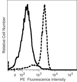
The 12F1 monoclonal antibody specifically binds to CD49b. CD49b is also known as the integrin α2 chain (VLA-α2), a member of the integrin family of extracellular matrix and cell-cell adhesion receptors. VLA-α2 is non-covalently associated with VLA-β1 chain (CD29) in the VLA-2 complex. α2 is expressed on numerous cell types including activated T cells, platelets, and long-term cultivated T cells. α2β1 is a well-established receptor for collagen. This antibody is useful for studies of integrin expression and function.
Development References (6)
-
Hemler ME, Kawaguchi S, Bodorova J. CD49b cluster repoty. In: Schlossman SF. Stuart F. Schlossman .. et al., ed. Leucocyte typing V : white cell differentiation antigens : proceedings of the fifth international workshop and conference held in Boston, USA, 3-7 November, 1993. Oxford: Oxford University Press; 1995:1615-1616.
-
Hemler ME. VLA proteins in the integrin family: structures, functions, and their role on leukocytes. Annu Rev Immunol. 1990; 8:365-400. (Biology). View Reference
-
Knapp W. W. Knapp .. et al., ed. Leucocyte typing IV : white cell differentiation antigens. Oxford New York: Oxford University Press; 1989:1-1182.
-
Pischel KD, Bluestein HG, Woods VL, Jr. Platelet glycoproteins Ia, Ic, and IIa are physicochemically indistinguishable from the very late activation antigens adhesion-related proteins of lymphocytes and other cell type. J Clin Invest. 1988; 81(2):505-513. (Clone-specific: Immunoprecipitation). View Reference
-
Pischel KD, Hemler ME, Huang C, Bluestein HG, Woods VL, Jr. Use of the monoclonal antibody 12F1 to characterize the differentiation antigen VLA-2. J Immunol. 1987; 138(1):226-233. (Immunogen: Flow cytometry, Immunoprecipitation). View Reference
-
Schlossman SF. Stuart F. Schlossman .. et al., ed. Leucocyte typing V : white cell differentiation antigens : proceedings of the fifth international workshop and conference held in Boston, USA, 3-7 November, 1993. Oxford: Oxford University Press; 1995.
Please refer to Support Documents for Quality Certificates
Global - Refer to manufacturer's instructions for use and related User Manuals and Technical data sheets before using this products as described
Comparisons, where applicable, are made against older BD Technology, manual methods or are general performance claims. Comparisons are not made against non-BD technologies, unless otherwise noted.
For Research Use Only. Not for use in diagnostic or therapeutic procedures.
Report a Site Issue
This form is intended to help us improve our website experience. For other support, please visit our Contact Us page.