BD IMag™ Cell Separation Magnet
(RUO)
Description
The BD IMag™ Cell Separation Magnet is a specialized plastic test-tube rack surrounding a strong permanent rare earth magnet. It holds up to six 12 x 75-mm or two 17 x 100 mm round-bottom test tubes in optimal position for magnetic separation of leukocytes labeled with BD IMag™ particles - DM. When tubes containing BD IMag™-labeled cell suspensions are placed in the Cell Separation Magnet, the magnetic field attracts the labeled cells to the adjacent walls of the tubes, allowing removal of the supernatant containing any unlabeled cells. When the tubes are subsequently removed from the Cell Separation Magnet, the labeled cells may be harvested. This simple system allows rapid and effective positive or negative selection of desired leukocyte populations.
The Cell Separation Magnet contains a strong permanent magnet that may affect or damage delicate instruments, electronic equipment, and magnetic recording devices. Therefore, persons wearing cardiac pacemakers should not handle the Cell Separation Magnet, and the Cell Separation Magnet should not be placed near laboratory instruments, monitors, computer discs, credit cards, video or audio cassettes, and watches. The Cell Separation Magnet has a very strong attraction for ferric metals, so it should not be placed on steel surfaces or near objects containing steel or iron.
Like all precision instruments containing plastics, the Cell Separation Magnet may be warped or damaged by exposure to excessive heat or UV light. If disinfection is required, it may be wiped with alcohol or bleach solutions. Do not boil, autoclave, or expose the Cell Separation Magnet to artificial UV light.
Recommended Assay Procedures
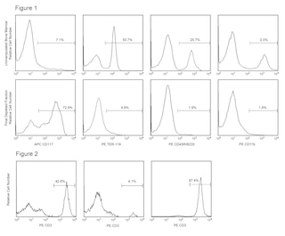
BD IMag™ particles - DM are optimal for use with the Cell Separation Magnet. A detailed protocol for both positive and negative selection is attached. BD IMag™ particles - MSC should not be used.
For negative selection of leukocyte subsets, antibodies are selected to label the cell population(s) that are not desired. Antibody-conjugated BD IMag™ particles - DM or biotinylated antibody plus Streptavidin BD IMag™ particles - DM are used to label the cell suspension, and the labeled cells are resuspended in 1X BD IMag™ buffer. The tube(s) containing the labeled cells are then placed in the Cell Separation Magnet, where the labeled cells are attracted to the magnet, and the desired cells remain suspended in the supernatant. With the tube(s) in the Cell Separation Magnet, the supernatant is harvested to obtain the desired leukocyte population.
For positive selection of leukocyte subsets, antibodies are selected to label the desired cell population. Antibody-conjugated BD IMag™ particles -DM or biotinylated antibody plus Streptavidin BD IMag™ particles - DM are used to label the cell suspension, and the labeled cells are resuspended in 1X BD IMag™ buffer. The tube(s) containing the labeled cells are then placed in the Cell Separation Magnet, where the desired cells are attracted to the magnet. With the tube(s) in the Cell Separation Magnet, the supernatant is removed to discard the undesired leukocytes. Then the tube is removed from the Cell Separation Magnet to allow harvesting of the desired cells.
CELL SEPARATION MAGNET PROTOCOL
This protocol is suitable for separation of single-cell suspensions of 1 x 10e7 to 2 x 10e8 leukocytes in 0.5 to 10 ml.
1. Label pairs of tubes appropriately to collect positive and negative fractions and place these on ice. Dilute BD IMag™ Buffer (10X) (Cat. No. 552362) 1:10 with sterile distilled water or prepare 1X BD IMag™ buffer from scratch (Phosphate Buffered Saline, 0.5% BSA, 2 mM EDTA, and 0.09% sodium azide) and place on ice.
Steps 2 - 5 should be performed for both positive and negative selection.
2. After the cells have been labeled with BD IMag™ particles - DM, adjust the cell concentration to 2 x 10e7 cells/ml with cold 1X BD IMag™ buffer.
3. Transfer the BD IMag™-labeled cell suspension to the positive-fraction collection tube(s):
- 12 x 75 mm tube: minimum volume of 0.5 ml and maximum volume of 3.5 ml
- 17 x 100 mm tube: minimum volume of 3.0 ml and maximum volume of 10 ml
4. Place the positive-fraction tube(s) into the appropriate tube holder(s) on the Cell Separation Magnet
- 12 x 75 mm tube: leave on the Cell Separation Magnet for 6 minutes
- 17 x 100 mm tube: leave on the Cell Separation Magnet for 10 minutes
5. With the positive-fraction tube(s) on the Cell Separation Magnet, remove the supernatant (negative fraction) using a glass pasteur pipette and transfer the negative fraction into the negative-fraction tube(s). Negative cell separation has now concluded. If positive selection is desired, continue to Step 6. If positive selection is not desired, go directly to Step 11.
6. Remove the positive-fraction tube(s) from the Cell Separation Magnet, and place on ice.
To further purify the positive fraction, we recommend the addition of Steps 7 - 10.
7. Add 1 ml of cold 1X BD IMag™ buffer to each positive-fraction tube. Resuspend cells by gently pipetting or vortexing. Place the tube(s) back on the Cell Separation Magnet.
- 12 x 75 mm tube: leave on the Cell Separation Magnet for 2 minutes
- 17 x 100 mm tube: leave on the Cell Separation Magnet for 5 minutes
8. Using a new glass pasteur pipette, remove supernatant and discard.
9. Repeat Steps 6, 7, and 8 one more time.
10. Remove the positive-fraction tube(s) from the Cell Separation Magnet and place it on ice. Add 1 ml of cold 1X BD IMag™ buffer to each positive-fraction tube. Resuspend cells by gently pipetting or vortexing. Positive-cell separation has now concluded.
11. Positive and negative fractions can now be resuspended in appropriate buffers for further downstream applications, including flow cytometry.
Product Notices
- Please refer to www.bdbiosciences.com/us/s/resources for technical protocols.
- BD IMag™ particles are prepared from carboxy-functionalized magnetic particles which are manufactured by Skold Technology and are licensed under US patent number 7,169,618.
Companion Products
Please refer to Support Documents for Quality Certificates
Global - Refer to manufacturer's instructions for use and related User Manuals and Technical data sheets before using this products as described
Comparisons, where applicable, are made against older BD Technology, manual methods or are general performance claims. Comparisons are not made against non-BD technologies, unless otherwise noted.
For Research Use Only. Not for use in diagnostic or therapeutic procedures.