

Preparation And Storage
Recommended Assay Procedures
MOUSE IgE ELISA PROTOCOL
I. Coat with Capture Antibody:
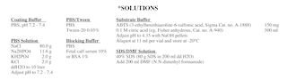
1. Dilute the purified anti-mouse IgE capture mAb (Cat. no. 553413, clone R35-72) to 2 µg/ml[a] in coating buffer (see solutions below).
Add 100 an enhanced protein-binding ELISA plate (eg. BD Falcon™ ELISA Plates, BD Labware Cat. no. 353279).
2. Shake plate to ensure all wells are covered by capture antibody solution.
3. Cover the plate and incubate for 1 hour at 37°C or overnight at 4°C.[b]
4. Wash the plate 3X with PBS/Tween (see solutions below). For each wash, wells are filled with 200 µl PBS/Tween and allowed to stand at
prior to aspirating or dumping. As a final step, tap plate on paper towels to remove excess buffer.
II. Blocking:
1. Block the plate with 200 µl blocking buffer (see solutions below) per well.
2. Cover the plate and incubate at room temperature for 30 minutes.
3. Wash the plate 3X with PBS/Tween, as in Section I, Step 4, of this protocol.
III. Apply Standards and Samples:
1. Leave column 1 as blank wells (ie, no antigen added, 100 µl per well blocking buffer only). Use columns 2 and 3 for duplicates
standard, 100 µl per well: dilute purified mouse IgE[a] standard (Cat. no. 557079, clone C38-2; or Cat. no. 553481,
mouse IgE[ b] standard (clone C48-2) in a series of 8 two-fold dilutions, in blocking buffer, starting at 0.5
the remaining columns to add samples at various dilutions in blocking buffer, 100 µl per well.
2. Cover the plate and incubate for at least 1 hour at room temperature or overnight at 4°C.[b]
3. Wash the plate 3X with PBS/Tween, as in Section I, Step 4, of this protocol.
IV. Incubation with Detection Antibody:
1. Dilute biotinylated anti-mouse IgE (Cat. no. 553419, clone R35-118) to 2 µg/ml[a] in blocking buffer. Add 100 µl per well.
2. Cover the plate and incubate at room temperature for 1 hour.
3. Wash the plate 6X with PBS/Tween, as in Section I, Step 4, of this protocol.
V. Add Avidin- or Streptavidin-Horseradish Peroxidase (Av-HRP or SAv-HRP):
1. Dilute Av-HRP (Cat. no. 554058) or SAv-HRP (Cat. no. 554066) 1:1000 in blocking buffer.[c] Add 100 µl per well.
2. Cover the plate and incubate at room temperature for 30 minutes.
3. Wash the plate 6X with PBS/Tween, as in Section I, Step 4, of this protocol.
VI. Add Substrate and Develop:
1. Thaw substrate (ABTS) buffer (see solutions below) within 20 minutes of use. Add 11 µl of 30% H2O2 (Sigma, Cat. no. H1009) to 11 ml
and vortex. Immediately add 100 µl per well and allow to develop at room temperature for 20 - 30 minutes. Color
stopped by adding 50 µl per well of SDS/DMF Solution (see solutions below) (optional).
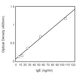
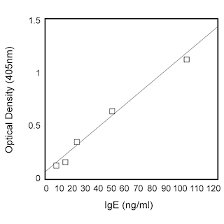
2. Read the plate at 405 nm.
NOTES
a. In most cases, coating the plate with primary mAb at 2 µg/ml, 100 µl per well and detecting with the biotinylated secondary mAb at 2 µg/ml,
yields a very satisfactory signal. However, for optimal signal, researchers should titrate each mAb over a range of concentrations (eg, 1 - 8
µg/ml).
b. Recommended incubation conditions for optimal sensitivity.
c. Streptavidin/Avidin-HRP conjugate from another supplier may be substituted and diluted according to the manufacturer's recommendation.
Product Notices
- Since applications vary, each investigator should titrate the reagent to obtain optimal results.
- Please refer to www.bdbiosciences.com/us/s/resources for technical protocols.
- Sodium azide is a reversible inhibitor of oxidative metabolism; therefore, antibody preparations containing this preservative agent must not be used in cell cultures nor injected into animals. Sodium azide may be removed by washing stained cells or plate-bound antibody or dialyzing soluble antibody in sodium azide-free buffer. Since endotoxin may also affect the results of functional studies, we recommend the NA/LE (No Azide/Low Endotoxin) antibody format, if available, for in vitro and in vivo use.
- Caution: Sodium azide yields highly toxic hydrazoic acid under acidic conditions. Dilute azide compounds in running water before discarding to avoid accumulation of potentially explosive deposits in plumbing.
Companion Products


The C48-2 antibody is specific for the hapten trinitrophenol (TNP).
Please refer to Support Documents for Quality Certificates
Global - Refer to manufacturer's instructions for use and related User Manuals and Technical data sheets before using this products as described
Comparisons, where applicable, are made against older BD Technology, manual methods or are general performance claims. Comparisons are not made against non-BD technologies, unless otherwise noted.
For Research Use Only. Not for use in diagnostic or therapeutic procedures.