Skip to main content Skip to navigation
APC Mouse Anti-Mouse Vβ 13 TCR
Multicolor flow cytometric analysis of Vβ 13 TCR expressed on mouse lymph node T cells.  C57BL/6 mouse lymph node cells were stained with PE Rat Anti-Mouse CD4 (Cat. No. 553048/553049) and PE Rat Anti-Mouse CD8a (Cat. No. 553032/553033) and either APC Mouse IgG1 κ Isotype Control (Cat. No. 554681, left panel) or an APC Mouse Anti-Mouse Vβ 13 TCR (Cat. No. 561542, right panel). Two-color flow cytometric dot plots showing the expression of Vβ 13 TCR (or Ig Isotype Control staining) versus CD4 and CD8 were derived from gated events with the forward and side light-scattering characteristics of viable lymphocytes. Flow cytometry was performed on a BD™ LSR II.
Product Details
Down Arrow Up Arrow


BD Pharmingen™
TCR V beta 13; TCR Vβ 13; TCR Vβ13; Vβ 13 T-cell Receptor
Mouse (QC Testing)
Mouse C57BR IgG1, κ
Not reported
Flow cytometry (Routinely Tested)
0.2 mg/ml
AB_10894393
Aqueous buffered solution containing ≤0.09% sodium azide.
RUO


Preparation And Storage

Store undiluted at 4°C and protected from prolonged exposure to light. Do not freeze. The monoclonal antibody was purified from tissue culture supernatant or ascites by affinity chromatography. The antibody was conjugated to APC under optimum conditions, and unconjugated antibody and free APC were removed.

Product Notices

  1. Since applications vary, each investigator should titrate the reagent to obtain optimal results.
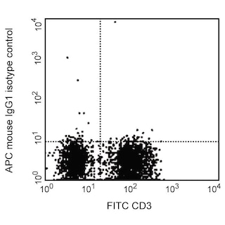
  2. An isotype control should be used at the same concentration as the antibody of interest.
  3. Caution: Sodium azide yields highly toxic hydrazoic acid under acidic conditions. Dilute azide compounds in running water before discarding to avoid accumulation of potentially explosive deposits in plumbing.
  4. This APC-conjugated reagent can be used in any flow cytometer equipped with a dye, HeNe, or red diode laser.
  5. For fluorochrome spectra and suitable instrument settings, please refer to our Multicolor Flow Cytometry web page at www.bdbiosciences.com/colors.
  6. Please refer to www.bdbiosciences.com/us/s/resources for technical protocols.
561542 Rev. 2
Antibody Details
Down Arrow Up Arrow
MR12-3

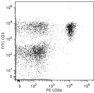
The MR12-3 monoclonal antibody specifically binds to the Vβ 13 T-cell Receptor (TCR) of mice having the b haplotype (e.g., A, AKR, BALB/c, CBA, C3H/He, C57BL, C58, DBA/1, DBA/2) of the Tcrb gene complex. The Tcrb-V13 gene locus is deleted in mice having the a (e.g., C57BR, C57L, SJL, SWR) or c (e.g., RIII) haplotype.

561542 Rev. 2
Format Details
Down Arrow Up Arrow
APC
Allophycocyanin (APC), is part of the BD family of phycobiliprotein dyes. This fluorochrome is a multimeric fluorescent phycobiliprotein with excitation maximum (Ex Max) of 651 nm and an emission maximum (Em Max) at 660 nm. APC is designed to be excited by the Red (627-640 nm) laser and detected using an optical filter centered near 660 nm (e.g., a 660/20 nm bandpass filter). Please ensure that your instrument’s configurations (lasers and optical filters) are appropriate for this dye.
altImg
APC
Red 627-640 nm
651 nm
660 nm
561542 Rev.2
Citations & References
Down Arrow Up Arrow

Development References (3)

  1. Behlke MA, Chou HS, Huppi K, Loh DY. Murine T-cell receptor mutants with deletions of beta-chain variable region genes. Proc Natl Acad Sci U S A. 1986; 83(3):767-771. (Biology). View Reference
  2. Chen D, Lee F, Cebra JJ, Rubin DH. Predominant T-cell receptor Vbeta usage of intraepithelial lymphocytes during the immune response to enteric reovirus infection.. J Virol. 1997; 71(5):3431-6. (Biology). View Reference
  3. Haqqi TM, Banerjee S, Anderson GD, David CS. RIII S/J (H-2r). An inbred mouse strain with a massive deletion of T cell receptor V beta genes. J Exp Med. 1989; 169(6):1903-1909. (Biology). View Reference
561542 Rev. 2

Please refer to Support Documents for Quality Certificates


Global - Refer to manufacturer's instructions for use and related User Manuals and Technical data sheets before using this products as described


Comparisons, where applicable, are made against older BD Technology, manual methods or are general performance claims.  Comparisons are not made against non-BD technologies, unless otherwise noted.

For Research Use Only. Not for use in diagnostic or therapeutic procedures.