Preparation And Storage
Recommended Assay Procedures
For optimal and reproducible results, BD Horizon Brilliant Stain Buffer should be used anytime two or more BD Horizon Brilliant dyes (including BD OptiBuild Brilliant reagents) are used in the same experiment. Fluorescent dye interactions may cause staining artifacts which may affect data interpretation. The BD Horizon Brilliant Stain Buffer was designed to minimize these interactions. More information can be found in the Technical Data Sheet of the BD Horizon Brilliant Stain Buffer (Cat. No. 563794).
Product Notices
- This antibody was developed for use in flow cytometry.
- The production process underwent stringent testing and validation to assure that it generates a high-quality conjugate with consistent performance and specific binding activity. However, verification testing has not been performed on all conjugate lots.
- Researchers should determine the optimal concentration of this reagent for their individual applications.
- An isotype control should be used at the same concentration as the antibody of interest.
- Caution: Sodium azide yields highly toxic hydrazoic acid under acidic conditions. Dilute azide compounds in running water before discarding to avoid accumulation of potentially explosive deposits in plumbing.
- For fluorochrome spectra and suitable instrument settings, please refer to our Multicolor Flow Cytometry web page at www.bdbiosciences.com/colors.
- Please refer to www.bdbiosciences.com/us/s/resources for technical protocols.
- BD Horizon Brilliant Stain Buffer is covered by one or more of the following US patents: 8,110,673; 8,158,444; 8,575,303; 8,354,239.
- BD Horizon Brilliant™ Violet 750 is covered by one or more of the following US patents: 8,158,444; 8,802,450; 8,575,303; 8,455,613; 8,227,187; 8,841,072; 8,110,673.
Companion Products


The AER-37 (CRA-1) monoclonal antibody specifically recognizes FcεRIα which is also known as the high affinity immunoglobulin epsilon receptor subunit alpha (Fc-epsilon RI-alpha). FcεRIα is a type I transmembrane glycoprotein that is encoded by FCER1A (Fc fragment of IgE receptor Ia) which belongs to the Ig gene superfamily. FcεRIα serves as the binding subunit for the Fc region of IgE. It forms part of a heterotetrameric, high-affinity IgE Fc receptor (FceR1/FcεR1) that includes signal transducing subunits, one β-chain (FcεRIβ encoded by MS4A2) and two disulfide-linked γ-subunits (FcεRIγ encoded by FCER1G). FcεRIα is normally expressed on basophils and mast cells and can also be expressed on some monocytes, Langerhans cells, dendritic cells, and eosinophils from allergic donors. FcεRIα plays a major role in allergic responses and in the presentation of allergens to the immune system. The AER-37 antibody reportedly does not compete with IgE for FceR1 binding.
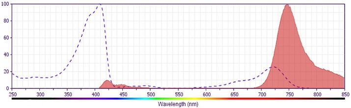
The antibody was conjugated to BD Horizon BV750 which is part of the BD Horizon Brilliant™ Violet family of dyes. This dye is a tandem fluorochrome of BD Horizon BV421 with an Ex Max of 405-nm and an acceptor dye with an Em Max at 750-nm. BD Horizon Brilliant BV750 can be excited by the violet laser (405 nm) and detected with a 750/30 nm filter with a 740 nm long pass. Due to spectral differences between labeled cells and beads, using BD™ CompBeads can result in incorrect spillover values when used with BD Horizon BV750 reagents. Therefore, the use of BD CompBeads or BD CompBeads Plus to determine spillover values for these reagents is not recommended.
Development References (6)
-
Cheng YX, Foster B, Holland SM, et al. CD2 identifies a monocyte subpopulation with immunoglobulin E-dependent, high-level expression of Fc epsilon RI.. Clin Exp Allergy. 2006; 36(11):1436-45. (Clone-specific: Flow cytometry). View Reference
-
Foster B, Metcalfe DD, Prussin C. Human dendritic cell 1 and dendritic cell 2 subsets express FcepsilonRI: correlation with serum IgE and allergic asthma.. J Allergy Clin Immunol. 2003; 112(6):1132-8. (Clone-specific: Flow cytometry). View Reference
-
Gounni AS, Lamkhioued B, Ochiai K, et al. High-affinity IgE receptor on eosinophils is involved in defence against parasites. Nature. 1994; 367(6459):183-186. (Biology). View Reference
-
Jürgens M, Wollenberg A, Hanau D, de la Salle H, Bieber T. Activation of human epidermal Langerhans cells by engagement of the high affinity receptor for IgE, Fc epsilon RI.. J Immunol. 1995; 155(11):5184-9. (Biology). View Reference
-
Maurer D, Fiebiger S, Ebner C, et al. Peripheral blood dendritic cells express Fc epsilon RI as a complex composed of Fc epsilon RI alpha- and Fc epsilon RI gamma-chains and can use this receptor for IgE-mediated allergen presentation.. J Immunol. 1996; 157(2):607-16. (Biology). View Reference
-
Takai T, Yuuki T, Iwamoto-Yasue N, Okumura K, Ra C. Epitope analysis and primary structures of variable regions of anti-human FcepsilonRI monoclonal antibodies, and expression of the chimeric antibodies fused with human constant regions.. Biosci Biotechnol Biochem. 2000; 64(9):1856-67. (Immunogen: Flow cytometry, Western blot). View Reference
Please refer to Support Documents for Quality Certificates
Global - Refer to manufacturer's instructions for use and related User Manuals and Technical data sheets before using this products as described
Comparisons, where applicable, are made against older BD Technology, manual methods or are general performance claims. Comparisons are not made against non-BD technologies, unless otherwise noted.
For Research Use Only. Not for use in diagnostic or therapeutic procedures.