Skip to main content Skip to navigation
BUV496 Streptavidin
Alert icon
This product is the replacement for [564666].
BUV496 Streptavidin
Flow cytometric analysis of CD45R/B220 expression on mouse splenocytes.   Mouse splenic leucocytes were either not stained (dashed line histogram) or were stained (solid line histogram) with Biotin Rat Anti-Mouse CD45R/B220 antibody (Cat. No. 553085/553086). The cells were then washed and further stained with BD Horizon™ BUV496 Streptavidin (Cat. No. 564666). Flow cytometric histograms were derived from gated events with the forward and side light-scatter characteristics of viable splenic leucocytes. Flow cytometric analysis was performed using a BD™ LSR II Flow Cytometer System.
Product Details
Down Arrow Up Arrow


BD Horizon™
Flow cytometry (Routinely Tested)
0.1 mg/ml
AB_2869599
Aqueous buffered solution containing BSA and ≤0.09% sodium azide.
RUO


Preparation And Storage

Store undiluted at 4°C and protected from prolonged exposure to light. Do not freeze. Streptavidin was conjugated with dye under optimum conditions, and unconjugated Streptavidin and free dye were removed

Recommended Assay Procedures

For optimal and reproducible results, BD Horizon Brilliant Stain Buffer should be used anytime two or more BD Horizon Brilliant dyes are used in the same experiment.  Fluorescent dye interactions may cause staining artifacts which may affect data interpretation.  The BD Horizon Brilliant Stain Buffer was designed to minimize these interactions.  More information can be found in the Technical Data Sheet of the BD Horizon Brilliant Stain Buffer (Cat. No. 563794/566349) or the BD Horizon Brilliant Stain Buffer Plus (Cat. No. 566385).

Note:  When using high concentrations of antibody, background binding of this dye to erythroid cell subsets (mature erythrocytes and precursors) has been observed.  For researchers studying these cell populations, or in cases where light scatter gating does not adequately exclude these cells from the analysis, this background may be an important factor to consider when selecting reagents for panel(s).

Product Notices

  1. Since applications vary, each investigator should titrate the reagent to obtain optimal results.
  2. Caution: Sodium azide yields highly toxic hydrazoic acid under acidic conditions. Dilute azide compounds in running water before discarding to avoid accumulation of potentially explosive deposits in plumbing.
  3. Source of all serum proteins is from USDA inspected abattoirs located in the United States.
  4. BD Horizon Brilliant Ultraviolet 496 is covered by one or more of the following US patents: 8,110,673; 8,158,444; 8,227,187; 8,575,303; and 8,354,239.
  5. For fluorochrome spectra and suitable instrument settings, please refer to our Multicolor Flow Cytometry web page at www.bdbiosciences.com/colors.
  6. BD Horizon Brilliant Stain Buffer is covered by one or more of the following US patents: 8,110,673; 8,158,444; 8,575,303; 8,354,239.
  7. Please refer to http://regdocs.bd.com to access safety data sheets (SDS).
  8. Please refer to www.bdbiosciences.com/us/s/resources for technical protocols.
Antibody Details
Down Arrow Up Arrow

Streptavidin is a non-glycosylated protein that is prepared chromatographically from the bacterium Streptomyces avidinii. Streptavidin homotetramers have a particularly high, non-covalent binding affinity for biotin. When conjugated with fluorochromes, streptavidin has been widely used with biotin-conjugated antibodies and other biotinylated specific-binding molecules (eg, recombinant proteins and lectins) to stain cells and tissues for subsequent multiparameter analysis by flow cytometry, fluorescence microscopy and imaging. Likewise, when conjugated with an enzyme (eg, Horseradish Peroxidase or Alkaline Phosphatase) and coupled with a colorimetric or luminescent substrate development system, streptavidin has found widespread use along with biotinylated antibodies in a number of applications including Western blot, ELISA, ELISPOT, immunocytochemistry and immunohistochemistry.

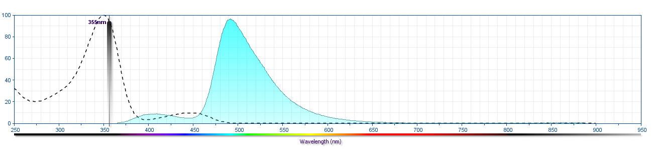
Streptavidin was conjugated to BD Horizon BUV496 which is part of the BD Horizon Brilliant™ Ultraviolet family of dyes. This dye is a tandem fluorochrome of BD Horizon BUV395 with an Ex Max of 348-nm and an acceptor dye with an Em Max at 496-nm. BD Horizon BUV496 can be excited by the ultraviolet laser (355 nm) and detected with a 515/30 nm filter with a 450LP. Due to the excitation of the acceptor dye by other laser lines, there may be significant spillover into the channel detecting BD Horizon V500 or BV510 (eg, 525/40-nm filter). However, the spillover can be corrected through compensation as with any other dye combination.

Format Details
Down Arrow Up Arrow
BUV496
The BD Horizon Brilliant™ Ultraviolet 496 (BUV496) Dye is part of the BD Horizon Brilliant™ Ultraviolet family of dyes. This tandem fluorochrome is comprised of a BUV395 donor with an excitation maximum (Ex Max) of 350-nm and an acceptor dye with an emission maximum (Em Max) at 496-nm. BUV496, driven by BD innovation, is designed to be excited by the ultraviolet laser (355-nm) and detected using an optical filter centered near 500-nm (e.g., 515/30-nm bandpass filter). The acceptor dye can be excited by the Violet (405-nm) laser resulting in cross-laser excitation and fluorescence spillover. Please ensure that your instrument’s configurations (lasers and optical filters) are appropriate for this dye.
altImg
BUV496
Ultraviolet 355 nm
350 nm
496 nm
Citations & References
Down Arrow Up Arrow

Development References (2)

  1. Diamandis EP, Christopoulos TK. The biotin-(strept)avidin system: principles and applications in biotechnology. Clin Chem. 1991; 37(5):625-636. (Biology). View Reference
  2. Shapiro HM. Practical flow cytometry, 4th ed.. Hoboken, N.J.: Wiley-Liss; 2003:1-681.

Please refer to Support Documents for Quality Certificates


Global - Refer to manufacturer's instructions for use and related User Manuals and Technical data sheets before using this products as described


Comparisons, where applicable, are made against older BD Technology, manual methods or are general performance claims.  Comparisons are not made against non-BD technologies, unless otherwise noted.

For Research Use Only. Not for use in diagnostic or therapeutic procedures.