Skip to main content Skip to navigation
BUV496 Mouse Anti-Human CD4
Alert icon
This on-demand reagent will be transitioning to a standard catalog equivalent in April 2024. Replacement SKU [569505] is available now. More Info #
BUV496 Mouse Anti-Human CD4
Multiparameter flow cytometric analysis using BD OptiBuild™ BUV496 Mouse Anti-Human CD4 antibody (Cat. No. 750591) on human peripheral blood. Flow cytometry was performed using a BD LSRFortessa™ X-20 Flow Cytometer System.
Product Details
Down Arrow Up Arrow


BD OptiBuild™
L3T4 ; T-cell surface antigen T4/Leu-3; W3/25 ; CD4 antigen (p55)
Human (Tested in Development)
Mouse BALB/c IgG1, κ
Human HPB-ALL Cell Line
Flow cytometry (Qualified)
0.2 mg/ml
AB_2874725
Aqueous buffered solution containing ≤0.09% sodium azide.
RUO


Preparation And Storage

Store undiluted at 4°C and protected from prolonged exposure to light. Do not freeze. The monoclonal antibody was purified from tissue culture supernatant or ascites by affinity chromatography. The antibody was conjugated with BD Horizon BUV496 under optimal conditions that minimize unconjugated dye and antibody.

Recommended Assay Procedures

For optimal and reproducible results, BD Horizon Brilliant Stain Buffer should be used anytime two or more BD Horizon Brilliant dyes (including BD OptiBuild Brilliant reagents) are used in the same experiment.  Fluorescent dye interactions may cause staining artifacts which may affect data interpretation.  The BD Horizon Brilliant Stain Buffer was designed to minimize these interactions.  More information can be found in the Technical Data Sheet of the BD Horizon Brilliant Stain Buffer (Cat. No. 563794).

Product Notices

  1. This antibody was developed for use in flow cytometry.
  2. The production process underwent stringent testing and validation to assure that it generates a high-quality conjugate with consistent performance and specific binding activity. However, verification testing has not been performed on all conjugate lots.
  3. Researchers should determine the optimal concentration of this reagent for their individual applications.
  4. An isotype control should be used at the same concentration as the antibody of interest.
  5. Caution: Sodium azide yields highly toxic hydrazoic acid under acidic conditions. Dilute azide compounds in running water before discarding to avoid accumulation of potentially explosive deposits in plumbing.
  6. For fluorochrome spectra and suitable instrument settings, please refer to our Multicolor Flow Cytometry web page at www.bdbiosciences.com/colors.
  7. Please refer to www.bdbiosciences.com/us/s/resources for technical protocols.
  8. BD Horizon Brilliant Stain Buffer is covered by one or more of the following US patents: 8,110,673; 8,158,444; 8,575,303; 8,354,239.
  9. BD Horizon Brilliant Ultraviolet 496 is covered by one or more of the following US patents: 8,110,673; 8,158,444; 8,227,187; 8,575,303; 8,354,239.
750591 Rev. 5
Antibody Details
Down Arrow Up Arrow
L200

The L200 monoclonal antibody specifically binds to the human form of the 56 kDa transmembrane glycoprotein, CD4, which is present on the T-helper/inducer subset of normal human donor peripheral blood lymphocytes. The L200 antibody also cross-reacts with a subset of CD3-positive peripheral blood lymphocytes, but not monocytes, of both Rhesus and Cynomolgus Macaque monkeys. Cross-reactivity on both lymphocytes and monocytes (weak) from baboons is also observed. CD4 distribution on lymphocytes is similar for both human and monkey cells, with the majority of CD4-positive lymphocytes being CD8-negative and lacking reactivity with antibodies to B- or NK-cell markers.

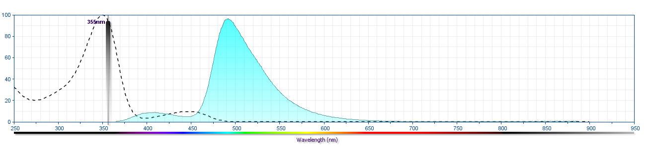
The antibody was conjugated to BD Horizon™ BUV496 which is part of the BD Horizon Brilliant™ Ultraviolet family of dyes. This dye is a tandem fluorochrome of BD Horizon BUV395 with an Ex Max of 348-nm and an acceptor dye with an Em Max at 496-nm. BD Horizon BUV496 can be excited by the ultraviolet laser (355 nm) and detected with a 515/30 nm filter with a 450LP. Due to the excitation of the acceptor dye by other laser lines, there may be significant spillover into the channel detecting BD Horizon V500 or BV510 (eg, 525/40-nm filter). However, the spillover can be corrected through compensation as with any other dye combination.

750591 Rev. 5
Format Details
Down Arrow Up Arrow
BUV496
The BD Horizon Brilliant™ Ultraviolet 496 (BUV496) Dye is part of the BD Horizon Brilliant™ Ultraviolet family of dyes. This tandem fluorochrome is comprised of a BUV395 donor with an excitation maximum (Ex Max) of 350-nm and an acceptor dye with an emission maximum (Em Max) at 496-nm. BUV496, driven by BD innovation, is designed to be excited by the ultraviolet laser (355-nm) and detected using an optical filter centered near 500-nm (e.g., 515/30-nm bandpass filter). The acceptor dye can be excited by the Violet (405-nm) laser resulting in cross-laser excitation and fluorescence spillover. Please ensure that your instrument’s configurations (lasers and optical filters) are appropriate for this dye.
altImg
BUV496
Ultraviolet 355 nm
350 nm
496 nm
750591 Rev.5
Citations & References
Down Arrow Up Arrow

Development References (11)

  1. Bleavins MR, Brott DA, Alvey JD, de la Iglesia FA. Flow cytometric characterization of lymphocyte subpopulations in the cynomolgus monkey (Macaca fascicularis). Vet Immunol Immunopathol. 1993; 37(1):1-13. (Biology). View Reference
  2. Giorgi JV, Hultin LE, Desrosiers RC. The immunopathogenesis of retroviral diseases: no immunophenotypic alterations in T, B, and NK cell subsets in SIVmac239-challenged rhesus macaques protected by SIV delta nef vaccination. J Med Primatol. 1996; 25(3):186-191. (Biology). View Reference
  3. Indzhiia LV, Yakovleva LA, Overbaugh J, et al. Baboon T cell lymphomas expressing the B cell-associated surface proteins CD40 and Bgp95. J Clin Invest. 1992; 12(3):225-236. (Biology). View Reference
  4. Jacobsen CN, Aasted B, Broe MK, Petersen JL. Reactivities of 20 anti-human monoclonal antibodies with leucocytes from ten different animal species. Vet Immunol Immunopathol. 1993; 39(4):461-466. (Biology). View Reference
  5. Lin G-X, Yang X, Hollemweguer E, et al. Cross-reactivity of CD antibodies in eight animal species. In: Mason D. David Mason .. et al., ed. Leucocyte typing VII : white cell differentiation antigens : proceedings of the Seventh International Workshop and Conference held in Harrogate, United Kingdom. Oxford: Oxford University Press; 2002:519-523.
  6. Powell JD, McClure HM, Anderson D, Fultz PN, Sell KW, Ahmed-Ansari A. Phenotypic and functional differences in NK and LAK cells in the peripheral blood of sooty mangabeys and rhesus macaques. Cell Immunol. 1989; 124(1):107-118. (Biology). View Reference
  7. Savary CA, Lotzova E, Jackson HJ, Jardine JH, Ang KK. Analysis of interleukin-2-activated killer cells of rhesus monkeys: striking resemblance to the human system. J Leukoc Biol. 1993; 54(4):307-313. (Biology). View Reference
  8. Schlossman SF. Stuart F. Schlossman .. et al., ed. Leucocyte typing V : white cell differentiation antigens : proceedings of the fifth international workshop and conference held in Boston, USA, 3-7 November, 1993. Oxford: Oxford University Press; 1995.
  9. Tryphonas H, Lacroix F, Hayward S, Izaguirre C, Parenteau M, Fournier J. Cell surface marker evaluation of infant Macaca monkey leukocytes in peripheral whole blood using simultaneous dual-color immunophenotypic analysis. J Med Primatol. 1996; 25(2):89-105. (Biology). View Reference
  10. Verdier F, Aujoulat M, Condevaux F, Descotes J. Determination of lymphocyte subsets and cytokine levels in cynomolgus monkeys. Toxicology. 1995; 105(1):81-90. (Biology). View Reference
  11. Wilson AD, Shooshtari M, Finerty S, Watkins P, Morgan AJ. Selection of monoclonal antibodies for the identification of lymphocyte surface antigens in the New World primate Saguinus oedipus oedipus (cotton top tamarin). J Immunol Methods. 1995; 178(2):195-200. (Biology). View Reference
View All (11) View Less
750591 Rev. 5

Please refer to Support Documents for Quality Certificates


Global - Refer to manufacturer's instructions for use and related User Manuals and Technical data sheets before using this products as described


Comparisons, where applicable, are made against older BD Technology, manual methods or are general performance claims.  Comparisons are not made against non-BD technologies, unless otherwise noted.

For Research Use Only. Not for use in diagnostic or therapeutic procedures.