Skip to main content Skip to navigation
PE Rat Anti-Mouse CD39
Flow cytometric analysis of CD39 expression on Mouse splenic lymphocytes. Balb/c mouse splenocytes were stained with FITC Rat Anti-Mouse CD4 (Cat. No. 553729, 557307, or 561828), APC Rat Anti-Mouse CD25 (Cat. No. 557192 or 561048), and PE Rat IgG2b, κ Isotype Control (Cat. No. 555848) or PE Rat Anti-Mouse CD39 antibody (Cat. No. 567104) at 0.5 μg/test. DAPI Solution (Cat. No. 564907) was added to cells right before analysis. Upper panels: Two-parameter flow cytometric pseudocolor plots showing correlated expression of CD39 (right) or Ig Isotype control (left) versus CD4 staining were derived from gated events with the forward and side light-scatter characteristics of viable (DAPI-negative) lymphocytes. Lower panels: Two-parameter flow cytometric pseudocolor plots showing correlated expression of CD39 (right) or Ig Isotype control (left) versus CD25 staining were derived from CD4-positive viable lymphocytes. Flow cytometry and data analysis were performed using a BD X-20 LSRFortessa™ Cell Analyzer System and FlowJo™ software. Data shown on this Technical Data Sheet are not lot specific.
Product Details
Down Arrow Up Arrow


BD Pharmingen™
ectonucleoside triphosphate diphosphohydrolase 1; Entpd1; ecto-ATP diphosphohydrolase 1; ecto-ATPDase 1; ecto-ATPase 1; Cd39; NTPDase 1
Mouse (QC Testing)
Rat F344, also known as Fischer, CDF IgG2b, κ
Mouse CD39 Transfected Cell Line
Flow cytometry (Routinely Tested)
0.2 mg/ml
12495
AB_2870021
Aqueous buffered solution containing ≤0.09% sodium azide.
RUO


Preparation And Storage

The monoclonal antibody was purified from tissue culture supernatant or ascites by affinity chromatography. The antibody was conjugated with R-PE under optimum conditions, and unconjugated antibody and free PE were removed. Store undiluted at 4°C and protected from prolonged exposure to light. Do not freeze.

Recommended Assay Procedures

BD® CompBeads can be used as surrogates to assess fluorescence spillover (compensation).  When fluorochrome conjugated antibodies are bound to BD® CompBeads, they have spectral properties very similar to cells.   However, for some fluorochromes there can be small differences in spectral emissions compared to cells, resulting in spillover values that differ when compared to biological controls.  It is strongly recommended that when using a reagent for the first time, users compare the spillover on cell and BD® CompBeads to ensure that BD® CompBeads are appropriate for your specific cellular application.

Product Notices

  1. Please refer to www.bdbiosciences.com/us/s/resources for technical protocols.
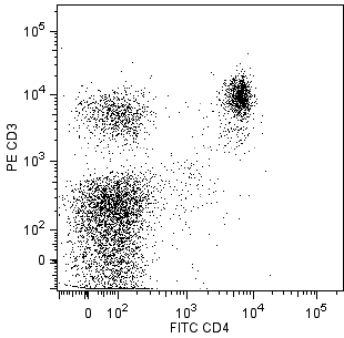
  2. Since applications vary, each investigator should titrate the reagent to obtain optimal results.
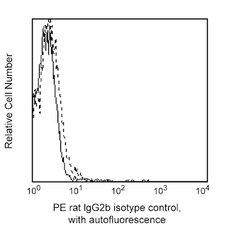
  3. An isotype control should be used at the same concentration as the antibody of interest.
  4. For fluorochrome spectra and suitable instrument settings, please refer to our Multicolor Flow Cytometry web page at www.bdbiosciences.com/colors.
  5. Caution: Sodium azide yields highly toxic hydrazoic acid under acidic conditions. Dilute azide compounds in running water before discarding to avoid accumulation of potentially explosive deposits in plumbing.
  6. Please refer to http://regdocs.bd.com to access safety data sheets (SDS).
567104 Rev. 2
Antibody Details
Down Arrow Up Arrow
Y23-1185

The Y23-1185 monoclonal antibody specifically recognizes mouse CD39, also known as ectonucleoside triphosphate diphosphohydrolase 1 (NTPDase 1), which is an enzyme on the surface of vascular endothelial cells, antigen presenting cells and activated immune cells. CD39 is encoded by ENTPD1 which belongs to the ectoenzyme family. The catalytic portion of the CD39 is extracellular, where it acts on extracellular nucleoside triphosphates and diphosphates, including ATP and ADP that are hydrolyzed into AMP. Through cell surface CD73 (Ecto-5'-nucleotidase), regulatory T cells can act on extracellular AMP to generate immunosuppressive adenosine. CD39 is involved in the control of the extracellular pool of phosphorylated nucleosides, the suppression of inflammation and immunity, and the regulation of platelet activation.

567104 Rev. 2
Format Details
Down Arrow Up Arrow
PE
R-Phycoerythrin (PE), is part of the BD family of Phycobiliprotein dyes. This fluorochrome is a multimeric fluorescent phycobiliprotein with excitation maximum (Ex Max) of 496 nm and 566 nm and an emission maximum (Em Max) at 576 nm. PE is designed to be excited by the Blue (488 nm), Green (532 nm) and Yellow-Green (561 nm) lasers and detected using an optical filter centered near 575 nm (e.g., a 575/26-nm bandpass filter). As PE is excited by multiple lasers, this can result in cross-laser excitation and fluorescence spillover on instruments with various combinations of Blue, Green, and Yellow-Green lasers. Please ensure that your instrument’s configurations (lasers and optical filters) are appropriate for this dye.
altImg
PE
Yellow-Green 488 nm, 532 nm, 561 nm
496 nm, 566 nm
576 nm
567104 Rev.2
Citations & References
Down Arrow Up Arrow

Development References (5)

  1. Allard D, Allard B, Stagg J. On the mechanism of anti-CD39 immune checkpoint therapy.. J Immunother Cancer. 2020; 8(1):e000186. (Biology). View Reference
  2. Borsellino G, Kleinewietfeld M, Di Mitri D, et al. Expression of ectonucleotidase CD39 by Foxp3+ Treg cells: hydrolysis of extracellular ATP and immune suppression.. Blood. 2007. (Biology). View Reference
  3. Mizumoto N, Kumamoto T, Robson SC, et al. CD39 is the dominant Langerhans cell-associated ecto-NTPDase: modulatory roles in inflammation and immune responsiveness.. Nat Med. 2002; 8(4):358-365. (Biology). View Reference
  4. Salmi M, Jalkanen S. Ectoenzymes controlling leukocyte traffic.. Eur J Immunol. 2012; 42(2):284-92. (Biology). View Reference
  5. Zhou Q, Yan J, Putheti P, et al. 2009; 9(10):2303-2311. (Biology). View Reference
View All (5) View Less
567104 Rev. 2

Please refer to Support Documents for Quality Certificates


Global - Refer to manufacturer's instructions for use and related User Manuals and Technical data sheets before using this products as described


Comparisons, where applicable, are made against older BD Technology, manual methods or are general performance claims.  Comparisons are not made against non-BD technologies, unless otherwise noted.

For Research Use Only. Not for use in diagnostic or therapeutic procedures.