-
Your selected country is
Middle East / Africa
- Change country/language
Old Browser
This page has been recently translated and is available in French now.
Looks like you're visiting us from {countryName}.
Would you like to stay on the current country site or be switched to your country?


Multiparameter flow cytometric analysis of Granzyme M expression in human peripheral blood leucocytes. Human peripheral blood cells were treated with BD Phosflow™ Lyse/Fix Buffer (Cat. No. 558049) to lyse erythrocytes and fix leucocytes. The leucocytes were washed and then stained in BD Perm/Wash™ Buffer (Cat. No. 554723) with either Alexa Fluor® 647 Mouse IgG1, κ Isotype Control (Cat. No. 565571; Left Plot) or Alexa Fluor® 647 Mouse Anti-Human Granzyme M antibody (Cat. No. 566996; Right Plot). A bivariate pseudocolor density plot showing the correlated expression of Granzyme M (or Ig Isotype control staining) versus side light-scatter (SSC) signals was derived from gated events with the forward and side light-scatter characteristics of intact leucocyte populations. Flow cytometric analysis was performed using a BD LSRFortessa™ X-20 Cell Analyzer System.

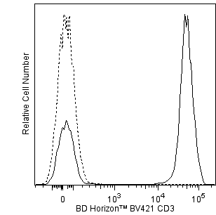
Two-color flow cytometric analysis of Granzyme M expression in human peripheral blood lymphocytes. Human peripheral blood cells were treated with BD Phosflow™ Lyse/Fix Buffer (Cat. No. 558049) to lyse erythrocytes and fix leucocytes. The leucocytes were washed and then stained in BD Perm/Wash™ Buffer (Cat. No. 554723) with BD Horizon™ BV421 Mouse anti-Human CD3 (Cat. No. 563798/563797) and either Alexa Fluor® 647 Mouse IgG1, κ Isotype Control (Cat. No. 565571; Left Plot) or Alexa Fluor® 647 Mouse Anti-Human Granzyme M antibody (Cat. No. 566996; Right Plot). A bivariate pseudocolor density plot showing the correlated expression of CD3 versus Granzyme M (or Ig Isotype control staining) was derived from gated events with the forward and side light-scatter characteristics of intact lymphocytes. Flow cytometric analysis was performed using a BD LSRFortessa™ X-20 Cell Analyzer System.


BD Pharmingen™ Alexa Fluor® 647 Mouse Anti-Human Granzyme M

BD Pharmingen™ Alexa Fluor® 647 Mouse Anti-Human Granzyme M

Regulatory Status Legend
Any use of products other than the permitted use without the express written authorization of Becton, Dickinson and Company is strictly prohibited.
Preparation And Storage
Recommended Assay Procedures
BD™ CompBeads can be used as surrogates to assess fluorescence spillover (Compensation). When fluorochrome conjugated antibodies are bound to BD CompBeads, they have spectral properties very similar to cells. However, for some fluorochromes there can be small differences in spectral emissions compared to cells, resulting in spillover values that differ when compared to biological controls. It is strongly recommended that when using a reagent for the first time, users compare the spillover on cells and BD CompBead to ensure that BD CompBeads are appropriate for your specific cellular application.
Product Notices
- This reagent has been pre-diluted for use at the recommended Volume per Test. We typically use 1 × 10^6 cells in a 100-µl experimental sample (a test).
- An isotype control should be used at the same concentration as the antibody of interest.
- Source of all serum proteins is from USDA inspected abattoirs located in the United States.
- Caution: Sodium azide yields highly toxic hydrazoic acid under acidic conditions. Dilute azide compounds in running water before discarding to avoid accumulation of potentially explosive deposits in plumbing.
- The Alexa Fluor®, Pacific Blue™, and Cascade Blue® dye antibody conjugates in this product are sold under license from Molecular Probes, Inc. for research use only, excluding use in combination with microarrays, or as analyte specific reagents. The Alexa Fluor® dyes (except for Alexa Fluor® 430), Pacific Blue™ dye, and Cascade Blue® dye are covered by pending and issued patents.
- Alexa Fluor® is a registered trademark of Molecular Probes, Inc., Eugene, OR.
- Alexa Fluor® 647 fluorochrome emission is collected at the same instrument settings as for allophycocyanin (APC).
- For fluorochrome spectra and suitable instrument settings, please refer to our Multicolor Flow Cytometry web page at www.bdbiosciences.com/colors.
- This product is provided under an intellectual property license between Life Technologies Corporation and BD Businesses. The purchase of this product conveys to the buyer the non-transferable right to use the purchased amount of the product and components of the product in research conducted by the buyer (whether the buyer is an academic or for-profit entity). The buyer cannot sell or otherwise transfer (a) this product (b) its components or (c) materials made using this product or its components to a third party or otherwise use this product or its components or materials made using this product or its components for Commercial Purposes. Commercial Purposes means any activity by a party for consideration and may include, but is not limited to: (1) use of the product or its components in manufacturing; (2) use of the product or its components to provide a service, information, or data; (3) use of the product or its components for therapeutic, diagnostic or prophylactic purposes; or (4) resale of the product or its components, whether or not such product or its components are resold for use in research. For information on purchasing a license to this product for any other use, contact Life Technologies Corporation, Cell Analysis Business Unit Business Development, 29851 Willow Creek Road, Eugene, OR 97402, USA, Tel: (541) 465-8300. Fax: (541) 335-0504.
- Please refer to http://regdocs.bd.com to access safety data sheets (SDS).
- Please refer to www.bdbiosciences.com/us/s/resources for technical protocols.
Companion Products


The 4B2G4 monoclonal antibody specifically recognizes Granzyme M which is also known as GrM, GzmM or Gzm M, as well as, Met-1 serine protease (Hu-Met-1) or Met-ase. Human Granzyme M belongs to a family of effector proteases that include five different granzymes that play key roles in immune responses to pathogens and transformed cells: Granzymes A, B, H, K and M. Granzyme M is a ~ 25-30 kDa neutral serine protease that is encoded by GZMM. Granzyme M participates in innate and adaptive immune responses as it is expressed in the cytotoxic granules of natural killer (NK) cells, NKT cells, cytotoxic αβ T cells and γδ T cells. Granzyme M participates in cell-mediated cytotoxicity by cleaving certain peptide substrates within target cells and promoting caspase activation and subsequent apoptosis. Granzyme M may also display non-cytotoxic functions including the inhibition of viral replication and the promotion of lipopolysaccharide (LPS)-mediated inflammatory responses.
Development References (7)
-
Anthony DA, Andrews DM, Chow M, et al. A role for granzyme M in TLR4-driven inflammation and endotoxicosis.. J Immunol. 2010; 185(3):1794-803. (Biology). View Reference
-
Bade B, Boettcher HE, Lohrmann J, et al. Differential expression of the granzymes A, K and M and perforin in human peripheral blood lymphocytes.. Int Immunol. 2005; 17(11):1419-28. (Clone-specific: Flow cytometry). View Reference
-
Bengsch B, Ohtani T, Herati RS, Bovenschen N, Chang KM, Wherry EJ. Deep immune profiling by mass cytometry links human T and NK cell differentiation and cytotoxic molecule expression patter. J Immunol Methods. 2018; 453:3-10. (Clone-specific). View Reference
-
Sayers TJ, Brooks AD, Ward JM, et al. The restricted expression of granzyme M in human lymphocytes.. J Immunol. 2001; 166(2):765-71. (Biology). View Reference
-
de Koning PJ, Tesselaar K, Bovenschen N, et al. The cytotoxic protease granzyme M is expressed by lymphocytes of both the innate and adaptive immune system.. Mol Immunol. 2010; 47(4):903-11. (Clone-specific: Flow cytometry). View Reference
-
de Poot SA, Bovenschen N. Granzyme M: behind enemy lines.. Cell Death Differ. 2014; 21(3):359-68. (Biology). View Reference
-
van Domselaar R, Philippen LE, Quadir R, Wiertz EJ, Kummer JA, Bovenschen N. Noncytotoxic inhibition of cytomegalovirus replication through NK cell protease granzyme M-mediated cleavage of viral phosphoprotein 71.. J Immunol. 2010; 185(12):7605-13. (Biology). View Reference
Please refer to Support Documents for Quality Certificates
Global - Refer to manufacturer's instructions for use and related User Manuals and Technical data sheets before using this products as described
Comparisons, where applicable, are made against older BD Technology, manual methods or are general performance claims. Comparisons are not made against non-BD technologies, unless otherwise noted.
For Research Use Only. Not for use in diagnostic or therapeutic procedures.
Report a Site Issue
This form is intended to help us improve our website experience. For other support, please visit our Contact Us page.