Skip to main content Skip to navigation
BV786 Mouse Anti-Human CD140a
Alert icon
If you're planning to order more than 10 BD OptiBuild™ Reagents, please contact your local sales representative to place this order. Contact Us #
Product Details
Down Arrow Up Arrow


BD OptiBuild™
PDGF Receptor α; PDGFRA; PDGFRα; PDGF-R-alpha; PDGFR2; PGFRA; RHEPDGFRA
Human (Tested in Development)
Mouse BALB/c IgG2a, κ
Human PDGFRα Transfected Cell Line
Flow cytometry (Qualified)
0.2 mg/ml
VI E031, BP229
5156
AB_2740957
Aqueous buffered solution containing ≤0.09% sodium azide.
RUO


Preparation And Storage

Store undiluted at 4°C and protected from prolonged exposure to light. Do not freeze. The monoclonal antibody was purified from tissue culture supernatant or ascites by affinity chromatography. The antibody was conjugated with BD Horizon BV786 under optimal conditions that minimize unconjugated dye and antibody.

Recommended Assay Procedures

For optimal and reproducible results, BD Horizon Brilliant Stain Buffer should be used anytime two or more BD Horizon Brilliant dyes (including BD OptiBuild Brilliant reagents) are used in the same experiment.  Fluorescent dye interactions may cause staining artifacts which may affect data interpretation.  The BD Horizon Brilliant Stain Buffer was designed to minimize these interactions.  More information can be found in the Technical Data Sheet of the BD Horizon Brilliant Stain Buffer (Cat. No. 563794).

Product Notices

  1. This antibody was developed for use in flow cytometry.
  2. The production process underwent stringent testing and validation to assure that it generates a high-quality conjugate with consistent performance and specific binding activity. However, verification testing has not been performed on all conjugate lots.
  3. Researchers should determine the optimal concentration of this reagent for their individual applications.
  4. An isotype control should be used at the same concentration as the antibody of interest.
  5. Caution: Sodium azide yields highly toxic hydrazoic acid under acidic conditions. Dilute azide compounds in running water before discarding to avoid accumulation of potentially explosive deposits in plumbing.
  6. For fluorochrome spectra and suitable instrument settings, please refer to our Multicolor Flow Cytometry web page at www.bdbiosciences.com/colors.
  7. Please refer to www.bdbiosciences.com/us/s/resources for technical protocols.
  8. BD Horizon Brilliant Stain Buffer is covered by one or more of the following US patents: 8,110,673; 8,158,444; 8,575,303; 8,354,239.
  9. BD Horizon Brilliant Violet 786 is covered by one or more of the following US patents: 8,110,673; 8,158,444; 8,227,187; 8,455,613; 8,575,303; 8,354,239.
  10. Cy is a trademark of GE Healthcare.
742669 Rev. 1
Antibody Details
Down Arrow Up Arrow
αR1

The αR1 monoclonal antibody specifically binds to the human platelet derived growth factor (PDGF) receptor α (PDGFRα), also known as CD140a. CD140a is a 170 kDa single transmembrane glycoprotein expressed on fibroblasts, smooth muscle cells, glial cells and chondrocytes. PDGF receptors α and β are single glycoproteins with intracellular tyrosine kinase domains. They are structurally similar to the M-CSF receptor and CD117 (c-kit). Their ligand, PDGF, is a mitogen for connective tissue cells and glial cells. PDGF plays a role in wound healing and it also acts as a chemoattractant for fibroblasts, smooth muscle cells, glial cells, monocytes and neutrophils. Functional PDGF is secreted in disulfide linked, homodimeric or heterodimeric forms comprised of A or B chains (PDGFAA, PDGF-BB or PDGF-AB). Binding of divalent PDGF induces receptor dimerization with three possible forms: αα, αβ, ββ. The PDGFRα subunit binds both PDGF A and B chains, whereas the PDGFRβ subunit binds only PDGF B chains. Although both receptor subunits can stimulate mitogenic responses, only the β subunit can induce chemotaxis. The αR1 antibody is specific for PDGFRα and does not crossreact with PDGFRβ. It immunoprecipitates human, monkey, rabbit, pig, dog and cat PDGFRα. It does not recognize hamster, rat or mouse PDGFRα.

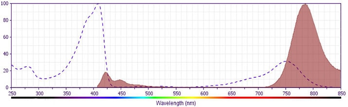
The antibody was conjugated to BD Horizon™ BV786 which is part of the BD Horizon Brilliant™ Violet family of dyes. This dye is a tandem fluorochrome of BD Horizon BV421 with an Ex Max of 405-nm and an acceptor dye with an Em Max at 786-nm.  BD Horizon BV786 can be excited by the violet laser and detected in a filter used to detect Cy™7-like dyes (eg, 780/60-nm filter).

742669 Rev. 1
Format Details
Down Arrow Up Arrow
BV786
The BD Horizon Brilliant Violet™ 786 (BV786) Dye is part of the BD Horizon Brilliant Violet™ family of dyes. This tandem fluorochrome is comprised of a BV421 donor with an Ex Max of 407-nm and an acceptor dye with an Em Max at 786-nm.  BV786, driven by BD innovation, is designed to be excited by the violet laser and detected using a filter, centered near 785 nm (e.g. 780/60 nm bandpass filter).  Please ensure that your instrument’s configurations (lasers and filters) are appropriate for this dye.
altImg
BV786
Violet 405 nm
407 nm
786 nm
742669 Rev.1
Citations & References
Down Arrow Up Arrow

Development References (5)

  1. Bazenet CE, Kazlauskas A. The PDGF receptor alpha subunit activates p21ras and triggers DNA synthesis without interacting with rasGAP. Oncogene. 1993; 9(2):517-525. (Biology). View Reference
  2. Callard R, Gearing A. Callard R, Gearing A. The Cytokine Facts Book. San Diego: Academic Press; 1994.
  3. Hart CE, Bowen-Pope DF. CD140a and b (PDGRα and β) Workshop Panel report. In: Kishimoto T. Tadamitsu Kishimoto .. et al., ed. Leucocyte typing VI : white cell differentiation antigens : proceedings of the sixth international workshop and conference held in Kobe, Japan, 10-14 November 1996. New York: Garland Pub.; 1997:739-741.
  4. Kitayama J, Springer TA. Endothelial Cell Blind Panel analysis: Overview and summary. In: Kishimoto T. Tadamitsu Kishimoto .. et al., ed. Leucocyte typing VI : white cell differentiation antigens : proceedings of the sixth international workshop and conference held in Kobe, Japan, 10-14 November 1996. New York: Garland Pub.; 1997:717-721.
  5. LaRochelle WJ, Jensen RA, Heidaran MA, et al. Inhibition of platelet-derived growth factor autocrine growth stimulation by a monoclonal antibody to the human alpha platelet-derived growth factor receptor. Cell Growth Differ. 1993; 4(7):547-553. (Immunogen: Flow cytometry, Functional assay, Immunoprecipitation). View Reference
View All (5) View Less
742669 Rev. 1

Please refer to Support Documents for Quality Certificates


Global - Refer to manufacturer's instructions for use and related User Manuals and Technical data sheets before using this products as described


Comparisons, where applicable, are made against older BD Technology, manual methods or are general performance claims.  Comparisons are not made against non-BD technologies, unless otherwise noted.

For Research Use Only. Not for use in diagnostic or therapeutic procedures.