

Preparation And Storage
Product Notices
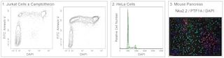
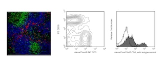
- This antibody has been developed for the immunofluorescence imaging application. However, the antibody is routinely QC tested by flow cytometric analysis. Researchers are encouraged to titrate the reagent for optimal performance.
- BD Horizon Brilliant Violet 480 is covered by one or more of the following US patents: 8,575,303; 8,354,239.
- ProLong® is a registered trademark of Thermo Fisher Scientific, Inc. Waltham, MA.
- Source of all serum proteins is from USDA inspected abattoirs located in the United States.
- Caution: Sodium azide yields highly toxic hydrazoic acid under acidic conditions. Dilute azide compounds in running water before discarding to avoid accumulation of potentially explosive deposits in plumbing.
- Please refer to www.bdbiosciences.com/us/s/resources for technical protocols.
Companion Products

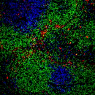
The T45-2342 monoclonal antibody recognizes the mouse F4/80 antigen which is also known as EGF-like module-containing mucin-like hormone receptor-like 1 (EMR1). F4/80 is a 160 kDa glycoprotein that belongs to the EGF-TM7 family of seven-transmembrane spanning cell surface molecules. It is expressed on the surface of granulocytes and a wide range of mature tissue macrophages including, Kupffer cells, splenic red pulp macrophages, microglia, gut lamina propria macrophages, and Langerhans cells. F4/80 expression has also been reported on subpopulations of dendritic cells. F4/80 expression is heterogeneous and may be increased during inflammatory responses as observed in various mouse models of colitis, diabetes and brain injury.
The antibody was conjugated to BD Horizon BV480 which is part of the BD Horizon Brilliant™ Violet family of dyes. This dye has been exclusively developed by BD Biosciences as an optimal dye for imaging applications. BD Horizon BV480 has an Ex Max at 436 nm and Em Max at 478 nm. The use of a mounting reagent (eg, ProLong® Gold) is highly recommended to maximize the photostability of BV480.
For confocal microscopy systems, a 440 nm laser is the optimal excitation source with optimal emission collection centered at 477 nm. BV480 fluorescence can also be excited well by 405 nm and 458 nm lasers.
For epifluorescence microscopes with broad spectrum excitation sources, the recommended excitation and emission filters are 445/20 nm and 485/20 nm bandpass filters, respectively. For specific multicolor imaging applications, the exact filter configurations should be optimized by the end user. For additional instrument/filter configuration information, please visit http://www.bdbiosciences.com/research/cellularimaging.

Development References (5)
-
Austyn JM., and Gordon S. F4/80, a monoclonal antibody directed specifically against the mouse macrophage. Eur J Immunol. 1981; 10:805-815. (Biology). View Reference
-
Gordon S, Hamann J, Lin HH, Stacey M. F4/80 and the related adhesion-GPCRs. Eur J Immunol. 2011; 41(9):2472-2476. (Biology). View Reference
-
Krüger T, Benke D, Eitner F, et al. Identification and functional characterization of dendritic cells in the healthy murine kidney and in experimental glomerulonephritis. J Am Soc Nephrol. 2004; 15(3):613-621. (Biology). View Reference
-
Leenen PJ, Radosević K, Voerman JS, et al. Heterogeneity of mouse spleen dendritic cells: in vivo phagocytic activity, expression of macrophage markers, and subpopulation turnover.. J Immunol. 1998; 160(5):2166-73. (Biology). View Reference
-
McKnight AJ, Macfarlane AJ, Dri P, Turley L, Willis AC, Gordon S. Molecular cloning of F4/80, a murine macrophage-restricted cell surface glycoprotein with Homology to the G-protein-linked transmembrane & hormone receptor family. J Biol Chem. 1996; 271:486. (Biology). View Reference
Please refer to Support Documents for Quality Certificates
Global - Refer to manufacturer's instructions for use and related User Manuals and Technical data sheets before using this products as described
Comparisons, where applicable, are made against older BD Technology, manual methods or are general performance claims. Comparisons are not made against non-BD technologies, unless otherwise noted.
For Research Use Only. Not for use in diagnostic or therapeutic procedures.