
Preparation And Storage
Recommended Assay Procedures
BD® CompBeads can be used as surrogates to assess fluorescence spillover (Compensation). When fluorochrome conjugated antibodies are bound to BD® CompBeads, they have spectral properties very similar to cells. However, for some fluorochromes there can be small differences in spectral emissions compared to cells, resulting in spillover values that differ when compared to biological controls. It is strongly recommended that when using a reagent for the first time, users compare the spillover on cells and BD® CompBeads to ensure that BD® CompBeads are appropriate for your specific cellular application.
For optimal and reproducible results, BD Horizon Brilliant™ Stain Buffer should be used anytime BD Horizon Brilliant™ dyes are used in a multicolor flow cytometry panel. Fluorescent dye interactions may cause staining artifacts which may affect data interpretation. The BD Horizon Brilliant™ Stain Buffer was designed to minimize these interactions. When BD Horizon Brilliant™ Stain Buffer is used in in the multicolor panel, it should also be used in the corresponding compensation controls for all dyes to achieve the most accurate compensation. For the most accurate compensation, compensation controls created with either cells or beads should be exposed to BD Horizon Brilliant™ Stain Buffer for the same length of time as the corresponding multicolor panel. More information can be found in the Technical Data Sheet of the BD Horizon Brilliant™ Stain Buffer (Cat. No. 563794/566349) or the BD Horizon Brilliant™ Stain Buffer Plus (Cat. No. 566385).
Product Notices
- This reagent has been pre-diluted for use at the recommended Volume per Test. We typically use 1 × 10^6 cells in a 100-µl experimental sample (a test).
- An isotype control should be used at the same concentration as the antibody of interest.
- Source of all serum proteins is from USDA inspected abattoirs located in the United States.
- Caution: Sodium azide yields highly toxic hydrazoic acid under acidic conditions. Dilute azide compounds in running water before discarding to avoid accumulation of potentially explosive deposits in plumbing.
- For fluorochrome spectra and suitable instrument settings, please refer to our Multicolor Flow Cytometry web page at www.bdbiosciences.com/colors.
- BD Horizon Brilliant Violet 786 is covered by one or more of the following US patents: 8,110,673; 8,158,444; 8,227,187; 8,455,613; 8,575,303; 8,354,239.
- BD Horizon Brilliant Stain Buffer is covered by one or more of the following US patents: 8,110,673; 8,158,444; 8,575,303; 8,354,239.
- Please refer to www.bdbiosciences.com/us/s/resources for technical protocols.
- Please refer to http://regdocs.bd.com to access safety data sheets (SDS).
- Cy is a trademark of Global Life Sciences Solutions Germany GmbH or an affiliate doing business as Cytiva.
Companion Products


The O33-782 monoclonal antibody specifically binds to Interleukin-17F (IL-17F). IL-17F is a member of the IL-17 family of cytokines. IL-17F is encoded by the IL17F gene located in chromosome 6 (location: 6p12). IL-17F is a proinflammatory cytokine that is produced by activated T cells including differentiated CD4+ T helper 17 (Th17) cells. Activated Th17 cells can express disulfide-linked IL-17F and IL-17A homodimers as well as IL-17A/IL-17F heterodimers. These IL-17 dimers act by binding to and signaling through IL-17 receptor complexes (IL-17R). IL-17R are comprised of transmembrane IL-17RA and IL-17-RC protein subunits that are expressed by a variety of target cells including epithelial and endothelial cells, keratinocytes, fibroblasts, and granulocytes. IL-17F can induce target cells to produce proinflammatory cytokines such as IL-1β, IL-6, G-CSF, GM-CSF, and TNF and chemokines including CXCL1/Gro-α, CXCL2/Gro-β, and CXCL8/IL-8 that attract and activate leukocytes, eg, neutrophils. Th17 and other IL-17F-producing cells play protective roles in the clearance of extracellular pathogens, including bacteria and fungi. IL-17F can also play adverse roles in inflammation associated with asthma and autoimmune diseases.
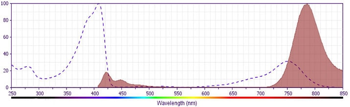
The antibody was conjugated to BD Horizon™ BV786 which is part of the BD Horizon Brilliant™ Violet family of dyes. This dye is a tandem fluorochrome of BD Horizon™ BV421 with an Ex Max of 405-nm and an acceptor dye with an Em Max at 786-nm. BD Horizon™ BV786 can be excited by the violet laser and detected in a filter used to detect Cy™7-like dyes (eg, 780/60-nm filter).
Development References (7)
-
Fouser LA, Wright JF, Dunussi-Joannopoulos K, Collins M. Th17 cytokines and their emerging roles in inflammation and autoimmunity. Immunol Rev. 2008; 226:87-102. (Biology). View Reference
-
Melton AC, Melrose J, Alajoki L, et al. Regulation of IL-17A production is distinct from IL-17F in a primary human cell co-culture model of T cell-mediated B cell activation. PLoS ONE. 2013; 8(3):e58966. (Clone-specific: Flow cytometry). View Reference
-
Shen F, Gaffen SL. Structure-function relationships in the IL-17 receptor: implications for signal transduction and therapy. Cytokine. 2008; 41(2):92-104. (Biology). View Reference
-
Starnes T, Robertson MJ, Sledge G, et al.. Cutting edge: IL-17F, a novel cytokine selectively expressed in activated T cells and monocytes, regulates angiogenesis and endothelial cell cytokine production. J Immunol. 2001; 167(8):4137-4140. (Biology). View Reference
-
Wang YH, Liu YJ. The IL-17 cytokine family and their role in allergic inflammation. Curr Opin Immunol. 2008; 20(6):697-702. (Biology). View Reference
-
Wright JF, Bennett F, Li B, et al. The human IL-17F/IL-17A heterodimeric cytokine signals through the IL-17RA/IL-17RC receptor complex. J Immunol. 2008; 181(4):2799-2805. (Biology). View Reference
-
Wright JF, Guo Y, Quazi A, et al. Identification of an interleukin 17F/17A heterodimer in activated human CD4+ T cells. J Biol Chem. 2007; 282(18):13447-13455. (Biology). View Reference
Please refer to Support Documents for Quality Certificates
Global - Refer to manufacturer's instructions for use and related User Manuals and Technical data sheets before using this products as described
Comparisons, where applicable, are made against older BD Technology, manual methods or are general performance claims. Comparisons are not made against non-BD technologies, unless otherwise noted.
For Research Use Only. Not for use in diagnostic or therapeutic procedures.