

Preparation And Storage
Product Notices
- This reagent has been pre-diluted for use at the recommended Volume per Test. We typically use 1 × 10^6 cells in a 100-µl experimental sample (a test).
- An isotype control should be used at the same concentration as the antibody of interest.
- Source of all serum proteins is from USDA inspected abattoirs located in the United States.
- Caution: Sodium azide yields highly toxic hydrazoic acid under acidic conditions. Dilute azide compounds in running water before discarding to avoid accumulation of potentially explosive deposits in plumbing.
- The Alexa Fluor®, Pacific Blue™, and Cascade Blue® dye antibody conjugates in this product are sold under license from Molecular Probes, Inc. for research use only, excluding use in combination with microarrays, or as analyte specific reagents. The Alexa Fluor® dyes (except for Alexa Fluor® 430), Pacific Blue™ dye, and Cascade Blue® dye are covered by pending and issued patents.
- Alexa Fluor® is a registered trademark of Molecular Probes, Inc., Eugene, OR.
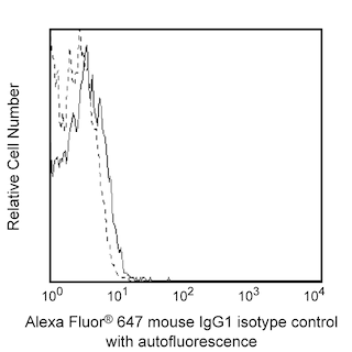
- Alexa Fluor® 647 fluorochrome emission is collected at the same instrument settings as for allophycocyanin (APC).
- For fluorochrome spectra and suitable instrument settings, please refer to our Multicolor Flow Cytometry web page at www.bdbiosciences.com/colors.
- Please refer to www.bdbiosciences.com/us/s/resources for technical protocols.
Companion Products


The 3D7 antibody reacts with CD131, the 120 kD common β chain (βc) which is shared with the receptor complexes for human granulocyte-macrophage colony stimulating factor (GM-CSFR), interleukin-3 (IL-3R) and interleukin-5 (IL-5R). Together with the α subunit of either the IL-3R (IL-3Rα), IL-5R (IL-5Rα), or GM- CSFR (GM-CSFRα), the common β chain forms high-affinity, signaling receptors for human IL-3, IL-5 and GM-CSF, respectively. Cell surface βc are expressed by a variety of different cell types including hematopoietic progenitor cells derived from pluripotent stem cells, monocytes, neutrophils, eosinophils, basophils, endothelial cells, fibroblasts, and Langerhans cells. The immunogen used to generate this hybridoma was cells co-transfected with expression vectors which contained cDNA for the human IL-3 α and β chains.
Development References (6)
-
CD64. In: Zola H. Leukocyte and stromal cell molecules : the CD markers. Hoboken, N.J.: Wiley-Liss; 2007:151.
-
Macardle PJ, Chen Z, Shih CY, et al. Characterization of human leucocytes bearing the IL-3 receptor. Cell Immunol. 1996; 168(1):59-68. (Clone-specific: Flow cytometry). View Reference
-
Miyajima A. CDw131 (interleukin 3 receptor β chain (common β)) Workshop Panel report. In: Kishimoto T. Tadamitsu Kishimoto .. et al., ed. Leucocyte typing VI : white cell differentiation antigens : proceedings of the sixth international workshop and conference held in Kobe, Japan, 10-14 November 1996. New York: Garland Pub.; 1997:859-861.
-
Woodcock JM, Zacharakis B, Plaetinck G. Three residues in the common beta chain of the human GM-CSF, IL-3 and IL-5 receptors are essential for GM-CSF and IL-5 but not IL-3 high affinity binding and interact with Glu21 of GM-CSF. EMBO J. 1994; 13(21):5176-5185. (Immunogen: Flow cytometry, Immunoprecipitation). View Reference
-
Zola H, Swart B, Banham A, et al. CD molecules 2006--human cell differentiation molecules.. J Immunol Methods. 2007; 319(1-2):1-5. (Clone-specific: Flow cytometry). View Reference
-
Zola H. Detection of cytokine receptors by flow cytometry. In: Coligan JE, Kruisbeek AM, Margulies DH, Shevach EM, Strober W, ed. Current Protocols in Immunology. New York: Green Publishing Associates and Wiley-Interscience; 1995:6.21.1-6.21.18.
Please refer to Support Documents for Quality Certificates
Global - Refer to manufacturer's instructions for use and related User Manuals and Technical data sheets before using this products as described
Comparisons, where applicable, are made against older BD Technology, manual methods or are general performance claims. Comparisons are not made against non-BD technologies, unless otherwise noted.
For Research Use Only. Not for use in diagnostic or therapeutic procedures.