
Preparation And Storage
Recommended Assay Procedures
Flow Cytometry: PE-Cy5 tandem fluorochromes have been reported to bind some classes of human macrophages and granulocytes via Fc receptors, and PE has been reported to bind to mouse B lymphocytes via Fc receptors. Preincubation of mouse leukocytes with Mouse BD Fc Block™ purified anti-mouse CD16/CD32 mAb 2.4G2 can reduce the non-specific binding of PE-Cy5-conjugated reagents to mouse B cells. However, PE-Cy5 conjugated reagents should not be used to stain splenocytes of SJL, NOD, and MRL mice as B lymphocytes and/or other leukocytes have been reported to non-specifically stain regardless of the use of Mouse BD Fc Block™. Reagents conjugated to PE, PerCP, PerCP-Cy5.5, APC, and APC-Cy7 tandem fluorochrome can be used on leukocytes from these mouse strains.
Product Notices
- Since applications vary, each investigator should titrate the reagent to obtain optimal results.
- Please refer to www.bdbiosciences.com/us/s/resources for technical protocols.
- Please observe the following precautions: Absorption of visible light can significantly alter the energy transfer occurring in any tandem fluorochrome conjugate; therefore, we recommend that special precautions be taken (such as wrapping vials, tubes, or racks in aluminum foil) to prevent exposure of conjugated reagents, including cells stained with those reagents, to room illumination.
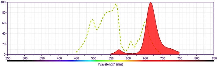
- For fluorochrome spectra and suitable instrument settings, please refer to our Multicolor Flow Cytometry web page at www.bdbiosciences.com/colors.
- PE-Cy5 is optimized for use with a single argon ion laser emitting 488-nm light. Because of the broad absorption spectrum of the PE-Cy5 tandem fluorochrome, extra care must be taken when using dual-laser cytometers which may directly excite both PE and Cy5™.
- PE-Cy5 is a tandem fluorochrome composed of R-phycoerythrin (PE), which is excited by the 488 nm light of an Argon ion laser and serves as an energy donor, coupled to the cyanine dye Cy5, which acts as an energy acceptor and fluoresces at 670 nm. BD Biosciences Pharmingen has maximized the fluorochrome energy transfer in PE-Cy5, thus maximizing its fluorescence emission intensity, minimizing residual emission from PE, and minimizing lot-to-lot variation.
- Cy is a trademark of Amersham Biosciences Limited. This conjugated product is sold under license to the following patents: US Patent Nos. 5,486,616; 5,569,587; 5,569,766; 5,627,027.
- This product is subject to proprietary rights of Amersham Biosciences Corp. and Carnegie Mellon University and made and sold under license from Amersham Biosciences Corp. This product is licensed for sale only for research. It is not licensed for any other use. If you require a commercial license to use this product and do not have one return this material, unopened to BD Biosciences, 10975 Torreyana Rd, San Diego, CA 92121 and any money paid for the material will be refunded.
- Caution: Sodium azide yields highly toxic hydrazoic acid under acidic conditions. Dilute azide compounds in running water before discarding to avoid accumulation of potentially explosive deposits in plumbing.
Companion Products


The RM4-5 monoclonal antibody specifically binds to the CD4 (L3T4) differentiation antigen expressed on most thymocytes, subpopulations of mature T lymphocytes (i.e., MHC class II-restricted T cells, including most T helper cells and immunosuppressive regulatory T cells), and a subset of NK-T cells. CD4 has also been reported to be detected on pluripotent hematopoietic stem cells, bone marrow myeloid and B-lymphocyte precursors, intrathymic lymphoid precursors, and a subset of splenic dendritic cells. CD4 has been reported to be expressed on the plasma membrane of mouse egg cells and is involved in adhesion of the egg to MHC class II-bearing sperm. CD4 is an antigen coreceptor on the T-cell surface which interacts with MHC class II molecules on antigen-presenting cells. It participates in T-cell activation through its association with the T-cell receptor complex and protein tyrosine kinase lck. Purified RM4-5 mAb has been reported to block the binding of FITC-conjugated anti-mouse CD4 clones GK1.5 and H129.19, but not the RM4-4 clone.
Development References (17)
-
Allman D, Li J, Hardy RR. Commitment to the B lymphoid lineage occurs before DH-JH recombination. J Exp Med. 1999; 189(4):735-740. (Biology). View Reference
-
Bendelac A. Mouse NK1+ T cells. Curr Opin Immunol. 1995; 7(3):367-374. (Biology). View Reference
-
Bierer BE, Sleckman BP, Ratnofsky SE, Burakoff SJ. The biologic roles of CD2, CD4, and CD8 in T-cell activation. Annu Rev Immunol. 1989; 7:579-599. (Biology). View Reference
-
Bosselut R, Zhang W, Ashe JM, Kopacz JL, Samelson LE, Singer A. Association of the adaptor molecule LAT with CD4 and CD8 coreceptors identifies a new coreceptor function in T cell receptor signal transduction. J Exp Med. 1999; 190(10):1517-1526. (Biology: Immunoprecipitation). View Reference
-
Frederickson GG, Basch RS. L3T4 antigen expression by hemopoietic precursor cells. J Exp Med. 1989; 169(4):1473-1478. (Biology). View Reference
-
Godfrey DI, Kennedy J, Mombaerts P, Tonegawa S, Zlotnik A. Onset of TCR-β gene rearrangement and role of TCR-β expression during CD3-CD4-CD8- thymocyte differentiation. J Immunol. 1994; 152(10):4783-4792. (Biology). View Reference
-
Guo MW, Watanabe T, Mori E, Mori T. Molecular structure and function of CD4 on murine egg plasma membrane. Zygote. 1995; 3(1):65-73. (Biology). View Reference
-
Janeway CA Jr. The T cell receptor as a multicomponent signalling machine: CD4/CD8 coreceptors and CD45 in T cell activation. Annu Rev Immunol. 1992; 10:645-674. (Biology). View Reference
-
Martin P, del Hoyo GM, Anjuere F, et al. Concept of lymphoid versus myeloid dendritic cell lineages revisited: both CD8alpha(-) and CD8alpha(+) dendritic cells are generated from CD4(low) lymphoid-committed precursors. Blood. 2000; 96(7):2511-2519. (Biology). View Reference
-
Nakamura T. Personal Communication. .
-
Shevach EM. Regulatory T cells in autoimmmunity. Annu Rev Immunol. 2000; 18:423-449. (Biology). View Reference
-
Takizawa F, Kinet JP, Adamczewski M. Binding of phycoerythrin and its conjugates to murine low affinity receptors for immunoglobulin G. J Immunol Methods. 1993; 162(2):269-272. (Methodology: Flow cytometry). View Reference
-
Waggoner AS, Ernst LA, Chen CH, Rechtenwald DJ. PE-CY5. A new fluorescent antibody label for three-color flow cytometry with a single laser. Ann N Y Acad Sci. 1993; 677:185-193. (Methodology: Flow cytometry). View Reference
-
Wineman JP, Gilmore GL, Gritzmacher C, Torbett BE, Muller-Sieburg CE. CD4 is expressed on murine pluripotent hematopoietic stem cells. Blood. 1992; 180(7):1717-1724. (Biology). View Reference
-
Wu L, Antica M, Johnson GR, Scollay R, Shortman K. Developmental potential of the earliest precursor cells from the adult mouse thymus. J Exp Med. 1991; 174(6):1617-1627. (Biology). View Reference
-
Wu L, Scollay R, Egerton M, Pearse M, Spangrude GJ, Shortman K. CD4 expressed on earliest T-lineage precursor cells in the adult murine thymus. Nature. 1991; 349(6304):71-74. (Biology). View Reference
-
van Vugt MJ, van den Herik-Oudijk IE, van de Winkle JG. Binding of PE-CY5 conjugates to the human high-affinity receptor for IgG (CD64). Blood. 1996; 88(6):2358-2361. (Methodology: Flow cytometry). View Reference
Please refer to Support Documents for Quality Certificates
Global - Refer to manufacturer's instructions for use and related User Manuals and Technical data sheets before using this products as described
Comparisons, where applicable, are made against older BD Technology, manual methods or are general performance claims. Comparisons are not made against non-BD technologies, unless otherwise noted.
For Research Use Only. Not for use in diagnostic or therapeutic procedures.