-
Your selected country is
Middle East / Africa
- Change country/language
Old Browser
This page has been recently translated and is available in French now.
Looks like you're visiting us from {countryName}.
Would you like to stay on the current country site or be switched to your country?


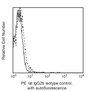
Flow cytometric analysis of CD104 expression on A431 cells, a human epidermal carcinoma cell line. A431 cells were stained with either PE Rat IgG2b, κ Isotype Control (Cat. No. 555848; dashed line histogram) or PE Rat Anti-Human CD104 (Cat. No. 555720; solid line histogram). Fluorescent histograms were derived from gated events with the side and forward light-scattering characteristics of viable A431 cells. Flow cytometry was performed on a BD FACScan™ system.


BD Pharmingen™ PE Rat Anti-Human CD104

Regulatory Status Legend
Any use of products other than the permitted use without the express written authorization of Becton, Dickinson and Company is strictly prohibited.
Preparation And Storage
Product Notices
- This reagent has been pre-diluted for use at the recommended Volume per Test. We typically use 1 × 10^6 cells in a 100-µl experimental sample (a test).
- An isotype control should be used at the same concentration as the antibody of interest.
- Source of all serum proteins is from USDA inspected abattoirs located in the United States.
- Caution: Sodium azide yields highly toxic hydrazoic acid under acidic conditions. Dilute azide compounds in running water before discarding to avoid accumulation of potentially explosive deposits in plumbing.
- For fluorochrome spectra and suitable instrument settings, please refer to our Multicolor Flow Cytometry web page at www.bdbiosciences.com/colors.
- Please refer to www.bdbiosciences.com/us/s/resources for technical protocols.
Companion Products


The 439-9B monoclonal antibody specifically recognizes CD104, integrin β4 chain, a 205 kDa transmembrane glycoprotein, which associates with CD49f (integrin α6 chain) to form the α6/β4 (CD49f/CD104) complex. It is expressed on epithelial cells, Schwann cells, and some tumor cells. The CD49f/CD104 complex is located in the hemidesmosomes of the epidermis, suggesting its role in epidermal cell-basement membrane adhesion. The clone 439-9B was clustered as CD104 at the fifth Human Leucocyte Differentiation Antigen International Workshop. It may be used for immunoprecipitation, immunoblotting and immunohistochemistry on frozen tissue sections.

Development References (7)
-
Falcioni R, Sacchi A, Resau J, Kennel SJ. Monoclonal antibody to human carcinoma-associated protein complex: quantitation in normal and tumor tissue.. Cancer Res. 1988; 48(4):816-21. (Immunogen). View Reference
-
Hemler ME, Crouse C, Sonnenberg A. Association of the VLA alpha 6 subunit with a novel protein. A possible alternative to the common VLA beta 1 subunit on certain cell lines. J Biol Chem. 1989; 264(11):6529-6535. (Biology). View Reference
-
Jones JC, Kurpakus MA, Cooper HM, Quaranta V. A function for the integrin alpha 6 beta 4 in the hemidesmosome. Cell Regul. 1991; 2(6):427-438. (Biology). View Reference
-
Mariani Costantini R, Falcioni R, Battista P, et al. Integrin (alpha 6/beta 4) expression in human lung cancer as monitored by specific monoclonal antibodies.. Cancer Res. 1990; 50(18):6107-12. (Biology). View Reference
-
Schlossman SF. Stuart F. Schlossman .. et al., ed. Leucocyte typing V : white cell differentiation antigens : proceedings of the fifth international workshop and conference held in Boston, USA, 3-7 November, 1993. Oxford: Oxford University Press; 1995.
-
Sonnenberg A, Calafat J, Janssen H, et al. Integrin alpha 6/beta 4 complex is located in hemidesmosomes, suggesting a major role in epidermal cell-basement membrane adhesion.. J Cell Biol. 1991; 113(4):907-17. (Biology). View Reference
-
Tamura RN, Rozzo C, Starr L, et al. Epithelial integrin alpha 6 beta 4: complete primary structure of alpha 6 and variant forms of beta 4.. J Cell Biol. 1990; 111(4):1593-604. (Biology). View Reference
Please refer to Support Documents for Quality Certificates
Global - Refer to manufacturer's instructions for use and related User Manuals and Technical data sheets before using this products as described
Comparisons, where applicable, are made against older BD Technology, manual methods or are general performance claims. Comparisons are not made against non-BD technologies, unless otherwise noted.
For Research Use Only. Not for use in diagnostic or therapeutic procedures.
Report a Site Issue
This form is intended to help us improve our website experience. For other support, please visit our Contact Us page.