Skip to main content Skip to navigation
PE Mouse anti-MEK1 (pS218)/MEK2 (pS222)
Flow cytometric and Western blot analyses of MEK1 (pS218)/MEK2 (pS222) expressed by Human and Mouse Cells. Human Cells. Panel 1.a. Human whole blood cells were either not stimulated (dashed line histogram) or were stimulated (solid line histogram) with Phorbol 12-Myristate 13-Acetate (PMA, Sigma, Cat. No. P8139; 400 nM, 15 min, 37°C). Cells were fixed in 1X BD Phosflow™ Lyse/Fix Buffer (Cat. No. 558049 for the 5X stock buffer; 10 min; 37˚C) and permeabilized in BD Phosflow™ Perm Buffer III (Cat. No. 558050; 30 min on ice). Cells were stained with BD Phosflow™ PE Mouse anti-MEK1 (pS218)/MEK2 (pS222) (Cat. No. 562458) antibody. Fluorescence histograms showing MEK1/2 (pS218/pS222) expression were generated for gated events with the forward and side-light scatter characteristics of intact peripheral blood lymphocytes (PBL). Flow cytometry was performed using a BD FACSCanto™ II Flow Cytometer System.   Panel 1.b. Cell lysates from 1 X 10^6 untreated (C) and PMA-treated (T) (50 nM PMA, 15 min, 37°C) human peripheral blood mononuclear cells (PBMC) were electrophoresed and Western blotted with Purified Mouse Anti-MEK1/2 (pS218) antibody (Cat. No. 562457; 2 µg/ml). Proteins were detected using HRP Goat Anti-Mouse Ig (Cat. No. 554002) and a chemiluminescent detection system. MEK1/2 (pS218/pS222) were identified as ~45 kDa bands. Mouse Cells. Panel 2.a. Mouse splenocytes were either not stimulated (dashed line histogram) or stimulated (solid line histogram) with PMA (50 nM, 15 min, 37°C). Cells were fixed, permeabilized, stained with BD Phosflow™ PE Mouse anti-MEK1 (pS218)/MEK2 (pS222) antibody and analyzed by flow cytometry as previously described.   Panel 2.b. Cell lysates from 1 X 10^6 untreated (C) and PMA-treated (T) (50 nM PMA, 15 min, 37°C) mouse splenocytes were analyzed by Western blot with Purified Mouse Anti-MEK1 (pS218)/MEK2 (pS222) antibody (2.0 µg/ml) as previously described. MEK1/2 (pS218/pS222) were identified as ~45 kDa bands.
Product Details
Down Arrow Up Arrow


BD Phosflow™
MAP2K1/2; MAPKK1/2; MAP kinase kinase 1/2; MAP2K1/2; PRKMK1/2
Human (QC Testing), Mouse (Tested in Development)
Mouse IgG1, κ
Phosphorylated Human MEK1 Peptide
Intracellular staining (flow cytometry) (Routinely Tested)
5 µl
AB_11153680
Aqueous buffered solution containing BSA and ≤0.09% sodium azide.
RUO


Preparation And Storage

Store undiluted at 4°C and protected from prolonged exposure to light. Do not freeze. The monoclonal antibody was purified from tissue culture supernatant or ascites by affinity chromatography. The antibody was conjugated with R-PE under optimum conditions, and unconjugated antibody and free PE were removed.

Recommended Assay Procedures

For more information please refer to the resources located on the BD Biosciences webpage: http://www.bdbiosciences.com/support/resources/index.jsp

Product Notices

  1. This reagent has been pre-diluted for use at the recommended Volume per Test. We typically use 1 × 10^6 cells in a 100-µl experimental sample (a test).
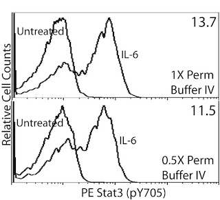
  2. An isotype control should be used at the same concentration as the antibody of interest.
  3. For fluorochrome spectra and suitable instrument settings, please refer to our Multicolor Flow Cytometry web page at www.bdbiosciences.com/colors.
  4. Please refer to www.bdbiosciences.com/us/s/resources for technical protocols.
  5. Caution: Sodium azide yields highly toxic hydrazoic acid under acidic conditions. Dilute azide compounds in running water before discarding to avoid accumulation of potentially explosive deposits in plumbing.
  6. Source of all serum proteins is from USDA inspected abattoirs located in the United States.
562458 Rev. 2
Antibody Details
Down Arrow Up Arrow
O24-836

The O24-836 monoclonal antibody specifically binds to the human MEK1 pS218 and MEK2 pS222 phosphorylated sites. MEK1 and MEK2 (MEK1/MEK2) are ~45 kDa dual-specificity protein kinases that are also known as MAPK/ERK Kinase 1 and 2 and MAP kinase kinases (aka, MAP2K1 and 2, MAPKK1 and 2, or MKK1 and 2).  MEK1 and MEK2 activation is dependent upon phosphorylation of certain amino acid residues including serine 218 (pS218) of MEK1 and serine 222 (pS222) of MEK2 by MAP kinase kinase kinases (MAP3Ks), such as by Raf kinases. MEK1/MEK2 can phosphorylate and thus activate the MAP (Mitogen-Activated Protein) kinases, ERK1 and ERK2. The ERK kinases are critically involved in multiple signal transduction pathways induced by growth factors, cytokines, and mitogens. These kinases regulate cellular growth, differentiation, survival, motility and angiogenesis. Dysregulated activation of MEK1/2 is observed in tumor cells. During antibody development, the purified O24-836 monoclonal antibody was found to detect phosphorylated MEK1/MEK2 by Western blot analysis of cellular lysates and by indirect immunofluorescent staining and flow cytometric analysis of fixed and permeabilized cells. This antibody crossreacts with phosphorylated MEK1/MEK2 expressed by mouse cells as tested by Western blot analysis and immunofluorescent staining and flow cytometric analysis.

562458 Rev. 2
Format Details
Down Arrow Up Arrow
PE
R-Phycoerythrin (PE), is part of the BD family of Phycobiliprotein dyes. This fluorochrome is a multimeric fluorescent phycobiliprotein with excitation maximum (Ex Max) of 496 nm and 566 nm and an emission maximum (Em Max) at 576 nm. PE is designed to be excited by the Blue (488 nm), Green (532 nm) and Yellow-Green (561 nm) lasers and detected using an optical filter centered near 575 nm (e.g., a 575/26-nm bandpass filter). As PE is excited by multiple lasers, this can result in cross-laser excitation and fluorescence spillover on instruments with various combinations of Blue, Green, and Yellow-Green lasers. Please ensure that your instrument’s configurations (lasers and optical filters) are appropriate for this dye.
altImg
PE
Yellow-Green 488 nm, 532 nm, 561 nm
496 nm, 566 nm
576 nm
562458 Rev.2
Citations & References
Down Arrow Up Arrow

Development References (5)

  1. Crews CM, Alessandrini A, Erikson RL. The primary structure of MEK, a protein kinase that phosphorylates the ERK gene product. Science. 1992; 258(5081):478-480. (Biology). View Reference
  2. Fremin C, Meloche S. From basic research to clinical development of MEK1/2 inhibitors for cancer therapy. J Hematol Oncol. 2010; 3(8):1-11. (Biology). View Reference
  3. Kolch W. Meaningful relationships: the regulation of the Ras/Raf/MEK/ERK pathway by protein interactions. Biochem J. 2000; 351:289-305. (Biology). View Reference
  4. Papin C, Eychene A, Brunet A, et al. B-Raf protein isoforms interact with and phosphorylate Mek-1 at serine residues 218 and 222. Oncogene. 1995; 10(8):1647-1651. (Biology). View Reference
  5. Yoon S, Seger R. The extracellular signal-regulated kinase: multiple substrates regulate diverse cellular functions. Growth Factors. 2006; 24(1):21-44. (Biology). View Reference
View All (5) View Less
562458 Rev. 2

Please refer to Support Documents for Quality Certificates


Global - Refer to manufacturer's instructions for use and related User Manuals and Technical data sheets before using this products as described


Comparisons, where applicable, are made against older BD Technology, manual methods or are general performance claims.  Comparisons are not made against non-BD technologies, unless otherwise noted.

For Research Use Only. Not for use in diagnostic or therapeutic procedures.